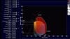
Margarita Revzin, M.D., MS, FSRU, FAIUM, associate professor of radiology and biomedical imaging, Yale University School of Medicine, abdominal and emergency imaging, radiologist, explains how different medical imaging modalities are used to image manifestations of the COVID-19 (SARS-CoV-2) virus in patients. She is the lead author on a two-part article in the RSNA journal Radiographics that provides a comprehensive overview of coronavirus imaging.
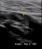
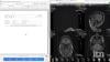
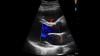
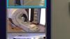
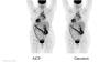
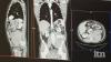
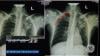
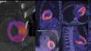
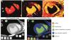
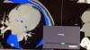
The articles offer numerous case images from X-ray, ultrasound, computed tomography (CT), and magnetic resonance imaging (MRI). Revzin also discusses some of the radiology presentations and complications of the virus and which modalities can best image these features. Here are links to the two articles:
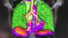
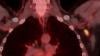
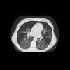
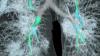
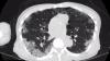
Manifestations of COVID-19, Part 1: Viral Pathogenesis and Pulmonary and Vascular System Complications.
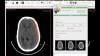
Multisystem Imaging Manifestations of COVID-19, Part 2: From Cardiac Complications to Pediatric Manifestations
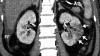
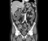
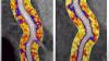
Although COVID-19 predominantly affects the respiratory system, other organs can also be involved. The authors of the articles said imaging plays an essential role in the diagnosis of all manifestations of the disease, as well as its related complications, and proper utilization and interpretation of imaging examinations is crucial. As the virus continues to spread, a comprehensive understanding of the diagnostic imaging hallmarks, imaging features, multisystemic involvement, and evolution of imaging findings is essential for effective patient management and treatment. Only a few articles had been published that comprehensively describe the multisystemic imaging manifestations of COVID-19 prior to this article series, published in the fall of 2020. The authors provide an inclusive system-by-system image-based review of this life-threatening and rapidly spreading infection. In part 1 of this article, the authors discuss general aspects of the disease, with an emphasis on virology, the pathophysiology of the virus, and clinical presentation of the disease. Part 2 focuses on key imaging features of COVID-19 that involve the cardiac, neurologic, abdominal, dermatologic and ocular, and musculoskeletal systems, as well as pediatric and pregnancy-related manifestations of the virus
Most of the images in the video are from the articles. Find more COVID medical imaging in the PHOTO GALLERY: How COVID-19 Appears on Medical Imaging.
Related Medical Imaging of COVID Content:
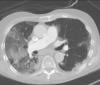
VIDEO: What Does COVID-19 Look Like in Lung CT Scans
PHOTO GALLERY: How COVID-19 Appears on Medical Imaging
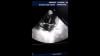
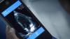
VIDEO: Imaging COVID-19 With Point-of-Care Ultrasound (POCUS) — Interview with Mike Stone, M.D.
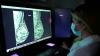
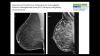
VIDEO: COVID Vaccine May Cause Enlarged Lymph Nodes on Mammograms — Interview with Constance "Connie" Lehman, M.D., Mass General Hospital
VIDEO: Use of Teleradiology During the COVID-19 Pandemic — Interview with John Kim, M.D.
VIDEO: Radiology Industry Responding to COVID-19 — Interview with Jeffrey Bundy, Ph.D.
CT in a Box Helps Rapidly Boost Imaging Capability at COVID Surge Hospitals
VIDEO: How China Leveraged Health IT to Combat COVID-19 — Interview with Jilan Liu, M.D.
Find more radiology related COVID content