June 2, 2026 — Results of an American College of Radiology-managed retrospective study involving 110,000 women presented at the 2026 ASCO Annual Meeting, shows that those who took GLP-1…
Breast Imaging
Women's health related to breast imaging, including mammography, breast MRI, ABUS, automated breast ultrasound, breast ultrasound, breast biopsy, PEM and positron emission mammography.
-

-

April 15, 2026 — QT Imaging Holdings, Inc.
-

April 1, 2026 — QT Imaging Holdings has released its latest image reconstruction software update, version 4.5.0.
-

March 30, 2026 — Each year, the Alumni Association at the University of Missouri-Kansas City, recognizes the achievements of outstanding alumni with an awards celebration.
-

March 2, 2026 — A collaborative modeling study found that adding biennial breast magnetic resonance imaging (MRI) to routine mammogram screening could avert more breast cancer deaths among women with extremely dense breasts and higher-than-average cancer risk.

News | MRI Breast
July 2, 2026 – Quibim has announced the European and UK launch of QP-Breast, its CE and UKCA-marked AI tool which ...
 July 02, 2026
News | Mammography
June 23, 2026 — Using artificial intelligence (AI), researchers found that image-based risk scores for breast cancer ...
June 24, 2026 
News | Women's Health
June 2, 2026 — Results of an American College of Radiology-managed retrospective study involving 110,000 women presented ...
June 02, 2026 Sponsored Content
Feature | Breast Imaging
Despite decades of progress in breast imaging, one challenge continues to test even the most skilled radiologists ...
October 24, 2025 
News | FDA
May 6, 2026 — Artera, the developer of multimodal artificial intelligence (MMAI)-based prognostic and predictive cancer ...
May 07, 2026 
News | Women's Health
April 16, 2026 – GE HealthCare has expanded its collaboration with DeepHealth, Inc., a wholly-owned subsidiary of RadNet ...
April 20, 2026 
News | Breast Imaging
April 15, 2026 — QT Imaging Holdings, Inc. has launched its QTI Imaging-Olea Viewer, developed in collaboration with ...
April 15, 2026 Sponsored Content
Feature | Breast Imaging
While most women understand the importance of health screenings, an estimated 72 million have missed or postponed a ...
December 03, 2024 
News | Breast Imaging
April 1, 2026 — QT Imaging Holdings has released its latest image reconstruction software update, version 4.5.0. This ...
April 02, 2026 
News | Breast Imaging
March 30, 2026 — Each year, the Alumni Association at the University of Missouri-Kansas City, recognizes the ...
March 31, 2026 
News | MRI Breast | Breast cancer, dense breast, MRI
March 2, 2026 — A collaborative modeling study found that adding biennial breast magnetic resonance imaging (MRI) to ...
March 20, 2026 Sponsored Content
Sponsored Content

Webinar | Mammography
The COVID-19 pandemic had a huge impact on the radiology community. Hospitals, doctors’ offices and clinics found ...
March 03, 2022  Clearance")
News | Breast Imaging
March 10, 2026 — QT Imaging Holdings has received U.S. Food and Drug Administration (FDA) 510(k) clearance for an ...
March 13, 2026 
News | Women's Health
March 4, 2026 — QT Imaging Holdings recently announced that Mary W. Yamashita, MD will serve as medical advisor to the ...
March 06, 2026 
News | Radiation Oncology
March 4, 2026 — Lunit has announced that 21 studies featuring its AI solutions will be presented at the European ...
March 05, 2026 Sponsored Content
Sponsored Content
Videos | Artificial Intelligence
Artificial Intelligence (AI) is becoming more common place in radiology practices, and emerging technologies are ...
November 11, 2020 
News | Women's Health
Feb.23, 2026 — The first clinical patient received a Clairity Breast cancer risk score, marking a historic milestone in ...
February 23, 2026 
News | Breast Biopsy Systems
Feb. 18, 2026 — Mammotome, a Danaher company, has introduced the Mammotome Prima MR Dual Vacuum-Assisted Breast Biopsy ...
February 18, 2026 
News | Breast Imaging
Feb. 16, 2026 — Rising demand for breast cancer screening and diagnostics is outpacing the supply of available breast ...
February 17, 2026 
News | FDA
Feb. 2, 2026 — Imagion Biosystems, Ltd. has submitted an Investigational New Drug (IND) application with the U.S. Food ...
February 02, 2026 
News | Breast Imaging | Washington University
Jan. 22, 2026 — In breast cancer, a biopsy is the only diagnostic procedure that can determine if a suspicious lump or ...
January 29, 2026 
News | Breast Imaging
Jan. 27, 2026 — QT Imaging and Olea Medical have announced plans to collaborate. This collaboration enhances QT Imaging ...
January 27, 2026 
News | Mammography
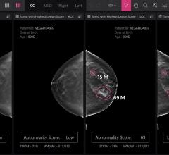
Jan. 16, 2026 — Vega Imaging Informatics has announced the successful curation of the world’s largest digital breast ...
January 19, 2026 
News | Breast Imaging
Jan. 14, 2026 — VizMark has received U.S. Food and Drug Administration FDA 510k clearance for VM1, a non-metal visual ...
January 19, 2026 © Copyright Wainscot Media. All Rights Reserved.
Subscribe Now