July 25, 2025 — Data in recent staffing surveys from the American Society of Radiologic Technologists show that vacancy rates for all medical imaging disciplines are above the rates reported in 2020, according to the 2025 ASRT Radiologic Sciences Staffing and Workplace Survey.
Coronavirus (COVID-19)
This page contains medical information for clinicians on the 2019 Novel Coronavirus (COVID-19, also called 2019-nCoV and now clinically SARS‐CoV‐2). This section includes articles on medical imaging of the virus for radiologists, new technologies being deployed to fight the virus and clinical information from various sources. Here are direct links for medical professionals to COVID-19 resources from the U.S. Food and Drug Administration (FDA), Centers for Disease Control (CDC) and the World Health Organization (WHO). Daily world-wide statistics on the coronavirus outbreak are available from the WHO Situations Reports. Centers for Medicare and Medicaid Services (CMS) frequently asked questions and answers (FAQs) for healthcare providers regarding Medicare payment for laboratory tests and other services related to the COVID-19
-

-

Postpandemic staffing shortages and increased volumes require radiologists to do more with less, exacerbating burnout.
-

This photo gallery shows the variety of radiological presentations of COVID-19 (SARS-CoV-2) in medical imaging, including computed tomography (CT), radiograph X-rays, ultrasound, echocardiograms and magnetic…
-
June 11, 2024 — A new study
-

March 21, 2024 — Artificial intelligence can spot COVID-19 in…

News | Radiology Imaging
July 25, 2025 — Data in recent staffing surveys from the American Society of Radiologic Technologists show that vacancy ...
 July 25, 2025
Videos | Coronavirus (COVID-19)
Postpandemic staffing shortages and increased volumes require radiologists to do more with less, exacerbating burnout ...
January 29, 2025 
News | Artificial Intelligence
June 11, 2024 — A new study led by researchers at Emory AI.Health, published in the Journal of Computers in Medicine and ...
June 11, 2024 Sponsored Content

Podcast | Enterprise Imaging
Members of the enterprise imaging technology community are facing significant changes in the market, due to the COVID-19 ...
March 17, 2021 
News | Coronavirus (COVID-19)
March 21, 2024 — Artificial intelligence can spot COVID-19 in lung ultrasound images much like facial recognition ...
March 21, 2024 
News | Coronavirus (COVID-19)
March 20, 2024 — SARS-CoV-2, the virus that causes COVID-19, can damage the heart even without directly infecting the ...
March 20, 2024 
News | Coronavirus (COVID-19)
November 22, 2023 — People with long COVID exhibit patterns of changes in the brain that are different from fully ...
November 22, 2023 Sponsored Content

Blog | Remote Viewing Systems
The COVID-19 pandemic accelerated a change that was already occurring in imaging-heavy practices across the country — ...
January 28, 2021 
News | Coronavirus (COVID-19)
November 20, 2023 — Using an image-guided minimally invasive procedure, researchers may be able to restore the sense of ...
November 20, 2023 
News | Cardiac Imaging
September 21, 2023 — Declines in cardiovascular procedure volumes observed early in the COVID-19 pandemic greatly ...
September 21, 2023 
News | SNMMI
September 18, 2023 — The Society of Nuclear Medicine and Molecular Imaging (SNMMI), as a professional society ...
September 18, 2023 Sponsored Content

Blog | Remote Viewing Systems
The coronavirus pandemic has changed the way radiologists read images and today facilities struggle to define a “new ...
August 04, 2020 
News | Artificial Intelligence
August 18, 2023 — Artificial intelligence (AI) is capturing the public imagination as the pace of innovation accelerates ...
August 18, 2023 
Feature | Information Technology | By Melinda Taschetta-Millane
Healthcare is constantly evolving, finding new ways to innovate and advance digital tools and technology. With this ...
July 13, 2023 
News | Contrast Media
July 3, 2023 — According to an accepted manuscript published in ARRS’ own American Journal of Roentgenology (AJR) ...
July 03, 2023 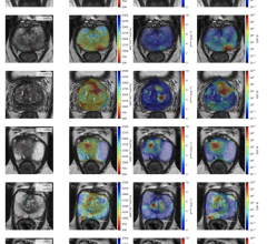
News | Coronavirus (COVID-19)
June 14, 2023 — A University of Waterloo engineer’s MRI invention reveals better than many existing imaging technologies ...
June 14, 2023 
News | Computed Tomography (CT)
May 17, 2023 — According to an accepted manuscript published in ARRS’ own American Journal of Roentgenology (AJR), SARS ...
May 17, 2023 
News | ACR
May 9, 2023 — The American College of Radiology (ACR) Economics and Health Policy Department has selected Ezequiel Silva ...
May 09, 2023 
News | Coronavirus (COVID-19)
March 13, 2023 — According to an accepted manuscript published in ARRS’ American Journal of Roentgenology (AJR) ...
March 13, 2023 
News | Coronavirus (COVID-19)
March 13, 2023 — The American College of Radiology (ACR) and the American College of Emergency Physicians (ACEP) ...
March 13, 2023 
News | Coronavirus (COVID-19)
March 10, 2023 — Researchers found evidence of heart muscle inflammation in a small number of patients with acute ...
March 10, 2023 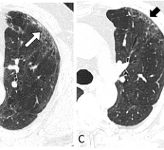
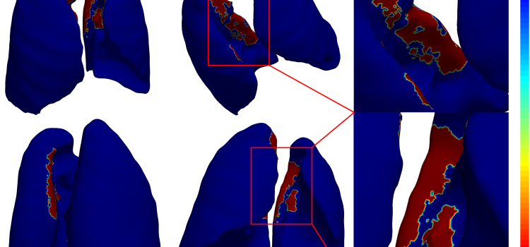
News | Lung Imaging
February 15, 2023 — Chest CT revealed persistent lung abnormalities in patients two years after COVID-19, according to a ...
February 15, 2023 
News | Coronavirus (COVID-19)
February 8, 2023 — New data published in the Journal of Clinical Oncology further quantify the vast lingering impact of ...
February 08, 2023 © Copyright Wainscot Media. All Rights Reserved.
Subscribe Now