
January 17, 2022 — The Society of Nuclear Medicine and Molecular Imaging (SNMMI) issued a statement on Jan. 14 regarding ...
Coronavirus (COVID-19)
This page contains medical information for clinicians on the 2019 Novel Coronavirus (COVID-19, also called 2019-nCoV and now clinically SARS‐CoV‐2). This section includes articles on medical imaging of the virus for radiologists, new technologies being deployed to fight the virus and clinical information from various sources. Here are direct links for medical professionals to COVID-19 resources from the U.S. Food and Drug Administration (FDA), Centers for Disease Control (CDC) and the World Health Organization (WHO). Daily world-wide statistics on the coronavirus outbreak are available from the WHO Situations Reports. Centers for Medicare and Medicaid Services (CMS) frequently asked questions and answers (FAQs) for healthcare providers regarding Medicare payment for laboratory tests and other services related to the COVID-19

News | Coronavirus (COVID-19)
January 14, 2022 — The COVID-19 pandemic took the world by storm in early 2020 and has become since then the leading ...
 January 14, 2022
News | Breast Imaging
January 11, 2022 — Less-experienced radiologists are more likely to recommend additional imaging for women undergoing ...
January 11, 2022 Sponsored Content

Podcast | Enterprise Imaging
Members of the enterprise imaging technology community are facing significant changes in the market, due to the COVID-19 ...
March 17, 2021 
News | Teleradiology
January 6, 2022 — Staffordshire University health experts claim there is an urgent need for global telehealth guidelines ...
January 06, 2022 
News | RSNA
January 5, 2022 — The Radiological Society of North America’s 107th Scientific Assembly and Annual Meeting (RSNA 2021) ...
January 05, 2022 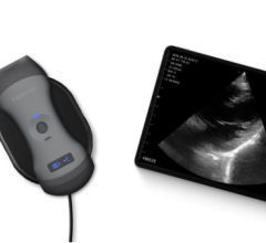
News | Ultrasound Imaging
January 4, 2022 — The future of ultrasound is happening — fast — and it doesn’t cost that much more than an iPhone. At ...
January 04, 2022 Sponsored Content

Blog | Remote Viewing Systems
The COVID-19 pandemic accelerated a change that was already occurring in imaging-heavy practices across the country — ...
January 28, 2021 
News | Information Technology
December 28, 2021 — Healthcare startups are constantly seeking to embrace emerging technologies to improve efficiency ...
December 28, 2021 
News | Teleradiology
December 27, 2021 — The "Global Radiology as a Service Market Size, Share & Trends Analysis Report by End User ...
December 27, 2021 
Feature | Radiology Imaging
This annual overview shows the top read content from ITNonline.com viewers throughout the year 2021. ITN had more than 2 ...
December 23, 2021 Sponsored Content

Blog | Remote Viewing Systems
The coronavirus pandemic has changed the way radiologists read images and today facilities struggle to define a “new ...
August 04, 2020 
News | Coronavirus (COVID-19)
December 23, 2021 — An interdisciplinary research team from the University of Göttingen and Hannover Medical School (MHH ...
December 23, 2021 
Feature | Radiology Business | By Melinda Taschetta-Millane
Here is a list of the Top 10 most read pieces of content on ITNonline.com from the month of December 2021. This is based ...
December 23, 2021 
Videos | Coronavirus (COVID-19)
Jean Jeudy, M.D., professor of radiology and vice chair of informatics at the University of Maryland School of Medicine ...
December 14, 2021 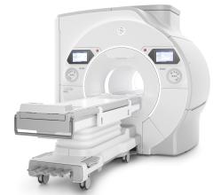
December 13, 2021 — GE Healthcare is proud to unveil SIGNA Hero[i], a new 3.0T magnetic resonance imaging (MRI) system ...
December 13, 2021 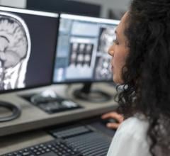
Videos | Teleradiology
Elizabeth Hawk, M.D., Ph.D., director of innovation Engagement at Rad Partners, a regional president for Matrix ...
December 10, 2021 
Feature | Radiology Imaging | By Melinda Taschetta-Millane
December 8, 2021 — Here is a list of the Top 10 most read pieces of content on ITNonline.com from the month of November ...
December 08, 2021 
News | Breast Imaging
December 7, 2021 — According to an open-access article in ARRS’ American Journal of Roentgenology (AJR), screening ...
December 07, 2021 
Videos | Coronavirus (COVID-19)
Kate Hanneman, M.D., MPH, FRCPC, director of cardiac imaging research JDMI, and the medical imaging site director at ...
December 06, 2021 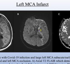
Videos | Coronavirus (COVID-19)
Scott Faro, M.D., professor of radiology and neurology and director, division of neuroradiology, head and neck, at ...
December 03, 2021 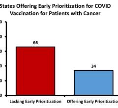
News | Coronavirus (COVID-19)
December 2, 2021 — Almost two-thirds of U.S. states failed to prioritize cancer patients for COVID-19 vaccinations ...
December 02, 2021 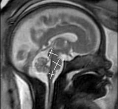
News | Coronavirus (COVID-19)
November 30, 2021 — COVID-19 of mild to moderate severity in pregnant women appears to have no effect on the brain of ...
November 30, 2021 © Copyright Wainscot Media. All Rights Reserved.
Subscribe Now