Jun. 24, 2025 — GE HealthCare has announced that the U.S. Food and Drug Administration (FDA) approved an updated label for its positron emission tomography (PET) imaging agent Vizamyl (flutemetamol F 18 injection) for beta-amyloid detection.
SNMMI
-

-

June 19, 2025 – dGenThera, Inc., a biotechnology company pioneering theranostic molecular pairs, and Nusano, a physics company transforming the production of radioisotopes, have announced the signing of a letter of intent to provide dGenThera with access to high‑purity astatine‑211 (At‑211
-
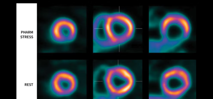
June 23, 2025 — GE HealthCare’s commitment to advancing precision care in cardiology through its molecular imaging solutions will be on display at the Society of Nuclear Medicine and Molecular Imaging (SNMMI) annual meeting in New Orleans, Louisiana.
-

June 23, 2025 — Serac Imaging Systems Ltd. and its clinical investigators from The Ohio State University Wexner Medical Center, U.S.
-
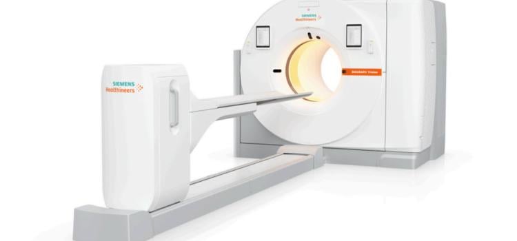
June 10, 2024 — Siemens Healthineers announces the Food and Drug Administration clearance of the Biograph Trinion, a high-performance, energy-efficient positron emission tomography/computed tomography (…

News | Nuclear Imaging
June 1, 2026 — At the 2026 Society of Nuclear Medicine and Molecular Imaging (SNMMI) annual meeting, GE HealthCare will ...
 June 02, 2026
June 1, 2026 — Serac Healthcare Ltd. has presented Phase 2 data showing that SPECT-CT imaging with the radiotracer 99mTc ...
June 01, 2026 
News | SNMMI
The Society of Nuclear Medicine and Molecular Imaging's (SNMMI) 2026 Annual Meeting will take place May 30–June 2 in Los ...
April 07, 2026 
News | PET Imaging
Jun. 24, 2025 — GE HealthCare has announced that the U.S. Food and Drug Administration (FDA) approved an updated label ...
June 24, 2025 
News | PET Imaging
June 19, 2025 – dGenThera, Inc., a biotechnology company pioneering theranostic molecular pairs, and Nusano, a physics ...
June 24, 2025 
News | PET-CT
June 23, 2025 — GE HealthCare’s commitment to advancing precision care in cardiology through its molecular imaging ...
June 23, 2025 
News | Nuclear Imaging
June 23, 2025 — Serac Imaging Systems Ltd. and its clinical investigators from The Ohio State University Wexner Medical ...
June 23, 2025 
News | Nuclear Imaging
June 20, 2024 — GE HealthCare joined the world’s top medical and academic institutions at the Society of Nuclear ...
June 20, 2024 
News | SNMMI
June 13, 2024 — The Society of Nuclear Medicine and Molecular Imaging (SNMMI) hosted more than 8,000 physicians ...
June 13, 2024 
News | Radiology Business
June 12, 2024 — Cathy Sue Cutler, PhD, FSNMMI, chair of the Isotope Research and Production Department at Brookhaven ...
June 12, 2024 
News | SNMMI
June 11, 2024 — Heather Jacene, MD, assistant chief of Nuclear Medicine and Molecular Imaging at Brigham and Women’s ...
June 11, 2024 
News | SNMMI
June 11, 2024 — The Society of Nuclear Medicine and Molecular Imaging recognized six new SNMMI Fellows during a plenary ...
June 11, 2024 
News | SPECT-CT
June 11, 2024 — A newly developed radiotracer can generate high quality and readily interpretable images of cardiac ...
June 11, 2024 
News | PET Imaging
June 11, 2024 — A new ultra-high-performance brain PET system allows for the direct measurement of brain nuclei as never ...
June 11, 2024 ![Jean-Luc C. Urbain, MD, PhD, FASNC, professor of Radiology/Nuclear Medicine and Medicine, has been named president-elect of the [Jean-Luc%20C.%20Urbain,%20MD,%20PhD,%20FASNC]Society of Nuclear Medicine and Molecular Imaging (SNMMI).](/sites/default/files/styles/feed_medium/public/Jean-Luc%20C.%20Urbain%2C%20MD%2C%20PhD%2C%20FASNC.jpg?itok=GtYJUnkI)
News | Radiology Business
June 10, 2024 — Jean-Luc C. Urbain, MD, PhD, FASNC, professor of Radiology/Nuclear Medicine and Medicine, has been named ...
June 10, 2024 
News | Radiology Business
June 10, 2024 — Carolyn J. Anderson, PhD, a trailblazer in nuclear medicine, has been named the 2024 recipient of the ...
June 10, 2024 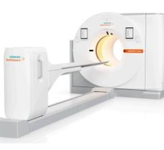
News | FDA
June 10, 2024 — Siemens Healthineers announces the Food and Drug Administration clearance of the Biograph Trinion, a ...
June 10, 2024 ![(A) PET images of [68Ga]Ga-DOTA-ZCAM241 uptake at baseline and 3, 7, and 12 days after injection as inflammatory arthritis developed in single representative individual mouse. Images are normalized to SUV of 0.5 for direct comparison between time points. (B) CD69 immunofluorescence Sytox (Thermo Fisher Scientific) staining of joints of representative animals during matching time points.](/sites/default/files/styles/feed_medium/public/PET%20Tracers.jpeg?itok=P5Di6MIe)
News | PET Imaging
February 9, 2024 — A novel PET imaging technique can noninvasively detect active inflammation in the body before ...
February 09, 2024 
News | SNMMI
February 6, 2024 — The Society of Nuclear Medicine and Molecular Imaging (SNMMI) held its 2024 SNMMI Mid-Winter Meeting ...
February 06, 2024 
News | PET Imaging
January 16, 2024 — The Society of Nuclear Medicine and Molecular Imaging (SNMMI) and the European Association of Nuclear ...
January 16, 2024 © Copyright Wainscot Media. All Rights Reserved.
Subscribe Now