June 10, 2026 — UTHealth Houston has launched a state-of-the-art PET/MRI imaging service, bringing together two advanced imaging technologies into a single integrated platform designed to enhance precision cancer care, support clinical research and expand opportunities in molecular imaging.
PET Imaging
Positron emission tomography (PET) is a nuclear imaging technology (also referred to as molecular imaging) that enables visualization of metabolic processes in the body. The basics of PET imaging is that the technique detects pairs of gamma rays emitted indirectly by a positron-emitting radionuclide (also called radiopharmaceuticals, radionuclides or radiotracer). The tracer is injected into a vein on a biologically active molecule, usually a sugar that is used for cellular energy. PET systems have sensitive detector panels to capture gamma ray emissions from inside the body and use software to plot to triangulate the source of the emissions, creating 3-D computed tomography images of the tracer concentrations within the body.
-

-

May 29, 2026 — GE HealthCare recently announced that its MIM KineticID modeling software1 is now 510(k) pending with the U.S. Food and Drug Administration.
-

Feb.
-

Jan. 8, 2026 — RefleXion Medical, an external-beam theranostic oncology company, has announced the U.S.
-

Nov. 18, 2025 — Siemens Healthineers positron emission tomography (PET) radiopharmaceutical companies PETNET Solutions Inc. and Advanced Accelerated Applications (AdAcAp) Molecular Imaging have been rebranded as Siemens Healthineers, effective immediately.

News | PET-MRI
June 10, 2026 — UTHealth Houston has launched a state-of-the-art PET/MRI imaging service, bringing together two advanced ...
 June 12, 2026
News | Nuclear Imaging
June 1, 2026 — At the 2026 Society of Nuclear Medicine and Molecular Imaging (SNMMI) annual meeting, GE HealthCare will ...
June 02, 2026 
News | PET Imaging
May 29, 2026 — GE HealthCare recently announced that its MIM KineticID modeling software1 is now 510(k) pending with the ...
May 29, 2026 Sponsored Content

Blog | PET Imaging
Digital technology is opening remarkable opportunities for clinical positron emission tomography (PET) about which ...
December 11, 2018 
News | PET Imaging
May 27, 2026 — A large multisite study of older people with cognitive impairment finds that Black and Hispanic people ...
May 29, 2026 
May 27, 2026 — Subtle Medical has received FDA clearance for its SubtleHD (PET), the company's next-generation AI ...
May 27, 2026 
May 7, 2026 — Bayer has announced positive topline results from the Phase III REVEAL study, an investigator-initiated ...
May 08, 2026 Sponsored Content

Blog | PET Imaging
Precision can have an enormous impact on patients. From diagnosis to patient monitoring (see “How Digital PET/CT Can ...
November 07, 2018 
News | PET Imaging
Catalyst MedTech Establishes Full Access Neurology Solution for Brain PET Implementation in the U.S.
March 25, 2026 — Catalyst MedTech, a provider of nuclear medicine and molecular imaging solutions, has announced it is ...
April 06, 2026 
News | PET Imaging
Feb. 19, 2026 — Positrigo, a Swiss based company developing nuclear medical devices to advance functional brain imaging ...
February 26, 2026 
News | Radiology Imaging
Feb. 12, 2026 — Siemens Healthineers and Mayo Clinic are expanding their strategic collaboration to enhance patient care ...
February 13, 2026 Sponsored Content

Podcast | PET Imaging
PET is getting ready to venture outside oncology, cardiology and mainstream neurology. High on the list of new clinical ...
November 07, 2018 
News | PET Imaging
Jan. 26, 2026 — Nuclidium, a clinical-stage radiopharmaceutical company developing a proprietary copper-based ...
January 27, 2026 
News | Radiation Oncology
Jan. 8, 2026 — RefleXion Medical, an external-beam theranostic oncology company, has announced the U.S. Food and Drug ...
January 13, 2026 
News | PET Imaging
Dec.16, 2025 — Three international nuclear medicine organizations — the SNMMI Clinical Trials Network (SNMMI-CTN), the ...
December 16, 2025 Sponsored Content
Blog | PET-CT
Analog is approximate. Digital is specific. Therein lies the fundamental difference between digital PET and its analog ...
October 17, 2018 
Nov. 18, 2025 — Siemens Healthineers positron emission tomography (PET) radiopharmaceutical companies PETNET Solutions ...
November 18, 2025 
News | PET Imaging
Oct. 22, 2025 — Vistim Labs has announced that its flagship product, Ceregram, is officially ready for launch in ...
October 22, 2025 
News | Radiology Imaging | UC San Diego Health
Oct. 16, 2025 — A strategic collaboration between UC San Diego Health and GE HealthCare will focus on bringing advanced ...
October 20, 2025 
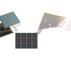
Sept. 20, 2025 — A promising new PET tracer can visualize a protein that is commonly overexpressed in triple-negative ...
September 18, 2025 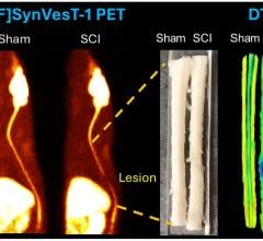
Sept. 11, 2025 — A new PET tracer can provide insights into how spinal cord injuries affect not only the spinal cord ...
September 12, 2025 
News | Artificial Intelligence
July 22, 2025 — GE HealthCare has topped a U.S. Food and Drug Administration (FDA) list of AI-enabled medical device ...
July 23, 2025 
News | PET Imaging
Jun. 24, 2025 — GE HealthCare has announced that the U.S. Food and Drug Administration (FDA) approved an updated label ...
June 24, 2025 
News | PET Imaging
June 19, 2025 – dGenThera, Inc., a biotechnology company pioneering theranostic molecular pairs, and Nusano, a physics ...
June 24, 2025 © Copyright Wainscot Media. All Rights Reserved.
Subscribe Now