Oct. 30, 2025 — Sirona Medical has received U.S. Food and Drug Administration (FDA) 510(k) clearance for its Sirona Advanced Imaging Suite, marking a significant regulatory milestone and the company's first Class II medical device designation.
PET-CT
PET-CT combines positron emission tomography (PET) detectors and computed tomography (CT) into one imaging system.
-

-

Aug. 5, 2025 — New Lantern has announced the launch of two specialized viewer modes: the Mammography Viewer Mode and PET/CT Viewer Mode.
-

June 23, 2025 — GE HealthCare’s commitment to advancing precision care in cardiology through its molecular imaging solutions will be on display at the Society of Nuclear Medicine and Molecular Imaging (SNMMI) annual meeting in New Orleans, Louisiana.
-

June 19, 2025 — Building on a collaboration that spans more than three decades, GE HealthCare has renewed its research collaboration with Stanford Medicine — with one of the key intentions being the development and research of innovative total body PET/CT technology.i This effort is ex
-
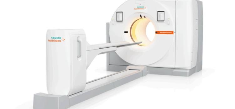
June 10, 2024 — Siemens Healthineers announces the Food and Drug Administration clearance of the Biograph Trinion, a high-performance, energy-efficient positron emission tomography/computed tomography (…

News | Radiology Imaging
April 20, 2026 — Bracco Imaging has announced a strategic alliance with NYU Langone Health to advance innovation in ...
 April 23, 2026
News | Radiology Business
March 31, 2026 — Radon Medical Imaging, a medical imaging equipment maintenance and repair services company, has has ...
March 31, 2026 
News | Remote Viewing Systems
Feb. 26, 2026 — DeepHealth, Inc., a provider of AI-powered health informatics and a wholly owned subsidiary of RadNet ...
February 27, 2026 Sponsored Content

Case Study | Enterprise Imaging
The healthcare industry faces many different types of obstacles in today’s challenging marketplace. Staff shortages ...
August 01, 2023 
News | PET-CT
Feb. 27, 2026 — A new targeted PET/CT tracer can detect treatment response in rheumatoid arthritis patients in as little ...
February 26, 2026 
News | FDA
Oct. 30, 2025 — Sirona Medical has received U.S. Food and Drug Administration (FDA) 510(k) clearance for its Sirona ...
October 30, 2025 
News | PET-CT
Oct. 6, 2025 — GE HealthCare has announced a strategic collaboration with Erasmus MC University Medical Center (Erasmus ...
October 17, 2025 Sponsored Content

Case Study | Enterprise Imaging
Reporting produces the tangible work product of diagnostic radiologists. Reports should reflect the expertise of the ...
October 30, 2022 
News | Prostate Cancer
Aug. 13, 2025 — A new study from Denmark shows for the first time that men with biochemically recurrent prostate cancer ...
August 15, 2025 
News | Mammography
Aug. 5, 2025 — New Lantern has announced the launch of two specialized viewer modes: the Mammography Viewer Mode and PET ...
August 05, 2025 
News | PET-CT
June 23, 2025 — GE HealthCare’s commitment to advancing precision care in cardiology through its molecular imaging ...
June 23, 2025 Sponsored Content

Blog | Radiology Imaging
People who live in rural America may deserve the same quality healthcare as anyone living in the U.S., but it is not ...
September 13, 2019 
News | PET-CT
June 19, 2025 — Building on a collaboration that spans more than three decades, GE HealthCare has renewed its research ...
June 19, 2025 
News | PET-CT
Nov. 25, 2024 — GE HealthCare has announced it is woking with Peter MacCallum Cancer Centre in Melbourne, Australia, to ...
November 25, 2024 
News | PET-CT
July 31, 2024 — In a head-to-head comparison with FDG PET/CT, FDG PET/MRI demonstrated comparable or superior diagnostic ...
July 31, 2024 Sponsored Content

News | PET-CT
Technological advancements in positron emission tomography/computed tomography (PET/CT) offer both clinicians and ...
February 06, 2019 
News | PET-CT
July 25, 2024 — Positron Corporation, a leading molecular imaging medical device company offering PET & PET-CT imaging ...
July 25, 2024 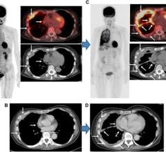
News | PET-CT
July 16, 2024 — A new research paper was published in Oncotarget's Volume 15 on June 20, 2024, titled, “Comparison of ...
July 16, 2024 
News | Prostate Cancer
July 2, 2024 — A new editorial paper was published in Oncoscience (Volume 11) on May 20, 2024, entitled, “Deep learning ...
July 02, 2024 
News | PET Imaging
June 18, 2024 — Positron Corporation, a leading molecular imaging medical device company offering PET and PET-CT ...
June 18, 2024 
News | PET Imaging
June 14, 2024 — Positron Corporation, a leading molecular imaging medical device company offering PET and PET-CT ...
June 14, 2024 
News | PET-CT
June 13, 2024 — Positron Corporation, a leading molecular imaging medical device company offering PET and PET-CT ...
June 13, 2024 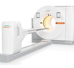
News | FDA
June 10, 2024 — Siemens Healthineers announces the Food and Drug Administration clearance of the Biograph Trinion, a ...
June 10, 2024 
News | Radiology Business
May 14, 2024 — University Hospitals (UH) and Siemens Healthineers announce a 10-year strategic alliance that builds on ...
May 14, 2024 © Copyright Wainscot Media. All Rights Reserved.
Subscribe Now