July 14, 2026 — A team of crew members aboard a commercial spaceflight acquired the first diagnostic X-rays during an orbital flight. Results of the mission were published recently in Radiology, a journal of the Radiological Society of North America (RSNA). “It’s been a dream for aerospace medicine
-

-

July 10, 2026 — A new study led by researchers at the Icahn School of Medicine at Mount Sinai found that although the overall number of radiation oncology clinics in the United States has appeared stable in recent years, many individual treatment centers have quietly closed while others have opened
-

Today’s radiology teams are faced with a wide range of growing complexities such as patient and procedure variabilities, workflow inefficiencies and the ongoing need to deliver consistent performance at scale. Bayer Radiology is addressing these challenges through protocol optimization and connected
-

Water-window X-rays allow researchers to visualize biological cells at high contrast without staining agents or other potentially damaging preparations. Multiple challenges, however, have prevented widespread adoption. Previously, the only way to produce water-window X-rays of varying energies was
-

June 9, 2026 — An investigator at the Icahn School of Medicine at Mount Sinai has received international recognition for MRI research that may help physicians identify patients at increased risk of chronic kidney disease (CKD) before they undergo surgery for kidney tumors. Mira Liu, PhD, a

News | Innovative Hospitals
Vanderbilt Health, Siemens Healthineers Partner to Deliver Advanced Technology, Elevate Patient Care
July 27, 2026 — Vanderbilt Health and Siemens Healthineers have entered into a multi-year, $87 million Value ...
 July 28, 2026
News | Computed Tomography (CT)
July 23, 2026 — HOPPR has introduced HOPPR EF Chest CT Narrative Model, a foundation model that processes 3D chest CT ...
July 27, 2026 
News | Pediatric Imaging
July 17, 2026 — In June, UW Health Kids began welcoming patients into a new state-of-the-art imaging suite dedicated to ...
July 23, 2026 Sponsored Content
Sponsored Content
Videos | Radiology Imaging
Today’s radiology teams are faced with a wide range of growing complexities such as patient and procedure variabilities ...
June 24, 2026 
News | ASTRO
July 22, 2026 — The American Society for Radiation Oncology (ASTRO) has announced the 39 members who will receive the ...
July 23, 2026 
July 21, 2026 — Fujifilm Sonosite has launched Sonosite iLOOK, a compact ultrasound solution designed specifically for ...
July 22, 2026 
News | RSNA
July 15, 2026 — The Radiological Society of North America has announced its lineup of plenary speaker for RSNA 2026 ...
July 21, 2026 Sponsored Content

Feature | Information Technology
AT A GLANCE Organization: Expert Radiology Management Services, LLC Specialty: Subspecialty teleradiology — neuro and ...
May 01, 2026 
News | Radiology Imaging
July 20, 2026 — Bracco Imaging has earned the 2026 EcoVadis Platinum Medal, the highest level of recognition awarded by ...
July 21, 2026 
News | Information Technology
July 20, 2026 — GE HealthCare has introduced MIM Anyware, a remote access platform that provides secure, healthcare ...
July 20, 2026 
News | Clinical Trials
July 15, 2026 — Varian, the cancer-care business of Siemens Healthineers, has published the results of its FAST-021 ...
July 17, 2026 Sponsored Content
Sponsored Content
Videos | Radiology Business
Radiology departments have many different needs and face a wide variety of challenges that can impact their departments ...
November 11, 2025 
News | Radiation Oncology
July 16, 2026 — Raidium has announced the U.S. launch of Raidium Read (R.Read), applying its AI-native imaging solution ...
July 16, 2026 
Feature | Information Technology | Kyle Hardner
Artificial intelligence (AI) has enhanced diagnostic accuracy and improved triage in radiology. But far fewer tools ...
July 16, 2026 
News | ACR
July 15, 2026 — The American College of Radiology (ACR) recently issued a statement praising the inclusion of the ...
July 16, 2026 Sponsored Content
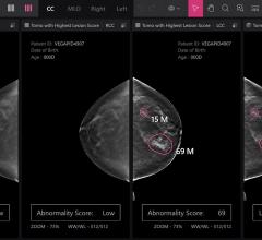
Feature | Breast Imaging
Despite decades of progress in breast imaging, one challenge continues to test even the most skilled radiologists ...
October 24, 2025 
News | Cardiac Imaging
July 8, 2026 — Conavi Medical Corp. has announced the publication of a case report in the Journal of the Society for ...
July 15, 2026 
News | PET Imaging
July 14, 2026 — New research is shedding new light on the biological basis of schizophrenia by directly measuring ...
July 15, 2026 
News | X-Ray
July 14, 2026 — A team of crew members aboard a commercial spaceflight acquired the first diagnostic X-rays during an ...
July 14, 2026 
News | Ultrasound Imaging
July 13, 2026 — Neal F. Kassell, MD, founder and chairman of the Focused Ultrasound Foundation, received the inaugural ...
July 13, 2026 
News | Radiation Oncology
July 10, 2026 — A new study led by researchers at the Icahn School of Medicine at Mount Sinai found that although the ...
July 10, 2026 
News | Neuro Imaging
July 9, 2026 — Viz.ai has announced a collaboration with Cortechs.ai to integrate Cortechs.ai's NeuroQuant and ...
July 10, 2026 
News | PACS
July 8, 2026 — Freeland Systems, a cloud PACS and clinical imaging software company, has launched its new customer ...
July 10, 2026 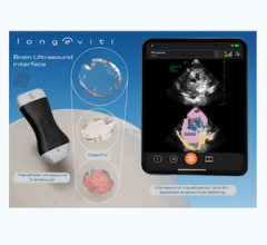
News | Ultrasound Imaging
July 7, 2026 — Longeviti Neuro Solutions has launched a new strategic initiative, ClearFit AI, a Brain Ultrasound ...
July 09, 2026 © Copyright Wainscot Media. All Rights Reserved.
Subscribe Now