News | Cardiac Imaging
April 28, 2026 — Abbott has received U.S. Food and Drug Administration (FDA) clearance and CE Mark for its next ...
 April 28, 2026
News | Radiology Business
April 24, 2026 — The 2026 vacancy rate for radiation therapists decreased to 11.4% and the vacancy rate for medical ...
April 24, 2026 
News | Contrast Agents
April 23, 2026 — On April 23, GE HealthCare announced the first patient has been dosed in the international, multi ...
April 23, 2026 Sponsored Content

Feature | Information Technology
AT A GLANCE Organization: Expert Radiology Management Services, LLC Specialty: Subspecialty teleradiology — neuro and ...
May 01, 2026 
News | Artificial Intelligence
April 20, 2026 — DeepTek, provider of the Augmento platform and deepc, the company behind deepcOS, have introduced a ...
April 23, 2026 
News | Radiology Imaging
April 20, 2026 — Bracco Imaging has announced a strategic alliance with NYU Langone Health to advance innovation in ...
April 23, 2026 
News | Women's Health
April 16, 2026 – GE HealthCare has expanded its collaboration with DeepHealth, Inc., a wholly-owned subsidiary of RadNet ...
April 20, 2026 Sponsored Content
Sponsored Content
Videos | Radiology Business
Radiology departments have many different needs and face a wide variety of challenges that can impact their departments ...
November 11, 2025 
News | FDA
April 16, 2026 — Royal Philips has received U.S. Food and Drug Administration 510(k) clearance for the Philips Spectral ...
April 20, 2026 
News | Breast Imaging
April 15, 2026 — QT Imaging Holdings, Inc. has launched its QTI Imaging-Olea Viewer, developed in collaboration with ...
April 15, 2026 
News | X-Ray
April 14, 2026 — KA Imaging is seeing continued adoption of its X-ray technology across new regions, with recent ...
April 15, 2026 Sponsored Content
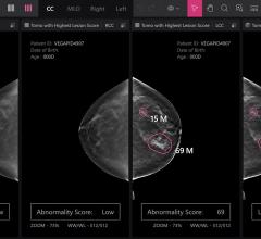
Feature | Breast Imaging
Despite decades of progress in breast imaging, one challenge continues to test even the most skilled radiologists ...
October 24, 2025 
News | Radiology Business
April 10, 2026 — The radiation therapy team at The Ohio State University Wexner Medical Center, The James Cancer ...
April 10, 2026 
News | Ultrasound Imaging
April 9, 2026 — GE HealthCare has announced a digital integration between the GE HealthCare bkActiv intraoperative ...
April 09, 2026 
News | Radiology Imaging
April 7, 2026 — Onvida Health and Siemens Healthineers have entered a 10-year Value Partnership¹ designed to bring the ...
April 09, 2026 Sponsored Content
Sponsored Content
Videos | Radiology Business
Bayer Radiology’s Barbara Ruhland and Thom Kinst discuss how radiology departments can address the many different ...
October 09, 2025 
April 8, 2026 — Alpha Imaging, a Radon Medical Imaging company, will display its latest advancement in image-guided ...
April 08, 2026 
News | Computed Tomography (CT)
April 2, 2026 — Nano-X Imaging Ltd. recently announced its U.S.-based subsidiary, Nanox Impact Inc., has signed a new ...
April 08, 2026 
News | SNMMI
The Society of Nuclear Medicine and Molecular Imaging's (SNMMI) 2026 Annual Meeting will take place May 30–June 2 in Los ...
April 07, 2026 
News | PET Imaging
Catalyst MedTech Establishes Full Access Neurology Solution for Brain PET Implementation in the U.S.
March 25, 2026 — Catalyst MedTech, a provider of nuclear medicine and molecular imaging solutions, has announced it is ...
April 06, 2026  Clearance to GE HealthCare's True Definition DL Software")
News | Computed Tomography (CT)
April 2, 2026 — GE HealthCare has received 510(k) clearance from the U.S. Food and Drug Administration (FDA) for True ...
April 03, 2026 
News | Teleradiology
April 1, 2026 — Premier Radiology Services has acquired Global Imaging Solutions (GLOBIS), a leading teleradiology group ...
April 03, 2026 
News | Breast Imaging
April 1, 2026 — QT Imaging Holdings has released its latest image reconstruction software update, version 4.5.0. This ...
April 02, 2026 
News | Ultrasound Imaging
March 30, 2026 — Butterfly Network, Inc. has received clearance from the U.S. Food and Drug Administration (FDA) for a ...
April 01, 2026 © Copyright Wainscot Media. All Rights Reserved.
Subscribe Now