
Micro-computed tomography provides non-destructive, qualitative and quantitative 3D analysis.
As scientists working in a range of life science and applied industrial applications seek deeper insights into complex new materials and biological structures, three-dimensional X-ray microscopy (3D XRM), also known as micro-computed tomography (micro-CT), is transforming imaging technology. The technique provides non-destructive, qualitative and quantitative 3D analysis of the microscopic world, giving insight into internal structures and shedding light on the mechanisms and success of fabrication.
Current instrumentation design has produced systems that are most often dedicated to a particular application and, as a result, the fixed architecture and components in those instruments have led to compromise – restricted sample size, field of view, analysis speed and fixed resolution, for example. For anyone looking to use the technology more flexibly, users have had to either deploy several instruments to perform different analyses depending on application or accept the need to swap out hardware elements and perform the necessary set up and recalibrations each time a change is made.
Recent innovations in imaging technology have enabled vendors to create modular, scalable and user-configurable systems, facilitating the development of versatile benchtop instruments. These radical advances have resulted in an imaging platform that allows a range of hardware components, required for different applications and analyses, to be mounted simultaneously on a single instrument. Such a “platform-based” approach can offer more flexibility, higher throughput, higher resolution imaging and faster time to result, compared to dedicated, floor standing systems.
An Advanced 3D XRM Configurable Platform
Having a modular platform that accommodates multiple configurations of key instrument components in one system means that laboratories can scale up their analysis functionality according to project needs over time or as new applications evolve. Importantly, to make a modular platform approach an attractive choice, there must be little or no compromise in performance, compared to previous generation instruments.
The potential benefits to a user of a configurable platform are clear – with no hardware modifications needed to switch the detector, sample charger or the calibrations required – reconfiguration of these components can be carried out by a straightforward software switch. This increases system uptime, optimizes lab productivity and avoids the need for instrument recalibration.
Important too, for the adoption of an accessible multi-scale benchtop microscopy solution is a straightforward workflow and control interface that can be operated by a user at any level, with minimal training. This requires pre-defined workflows that can be run by non-experts in multiple locations, in multiple local languages, with built-in safeguards to enable global deployment of processes and standard operating procedures from a central workflow-development source. In addition, a user management system that defines access levels per user group should be in place to ensure process consistency and data integrity.
A Versatile System
Examining the internal structure of biological and material samples without physical sectioning or other destructive preparation techniques preserves the sample integrity for retesting. For stable material analysis, 3D XRM allows a wide range of samples to be imaged in their native state, keeping sample preparation to a minimum. Where high throughput analysis is required, an automated changer can offer additional speed, but the best designed changers should retain the flexibility to manually insert a sample into the analysis flow.
Applications
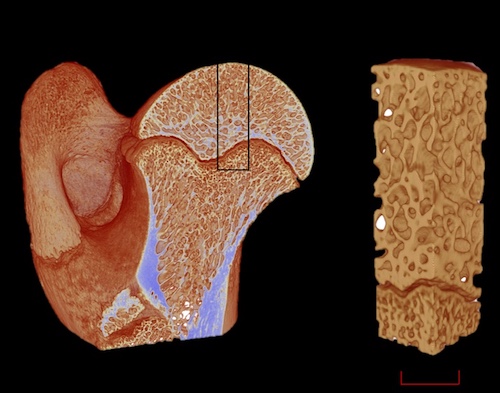
Osteology: Using 3D morphologic analysis extends understanding of the metabolic state and signaling responses of bone in preclinical imaging, helping to elucidate physiological and disease processes. Submicron imaging of mineralized bone opens the field of ultra-structural analysis, which includes visualization of blood vessel canals and osteocytes lacunae – features only visible at this level of resolution. The system operates at different resolutions depending on the sample and the experiment. Traditional methods require preparation, in the form of sectioning, that damage the sample. The 3D XRM system allows for non-destructive analysis of the internal microstructure of bone with high resolution and low noise active performance for ultrastructural analysis of whole bones.
Dentistry: The application of a platform-based solution for 3D XRM in evaluating the quality of dental fillings, detecting voids and assessing the fit of restorations contributes significantly to advancements in dental research and clinical practice.
A micro-CT system, equipped with a high-resolution detector, can provide detailed insights into the microstructural integrity of tooth structures and dental restorations and perform scans without destroying the samples, and for root canal preparation and filling techniques.
Among the materials science applications for 3D XRM are energy storage and pharmaceutical manufacturing.
Energy storage: A platform-based solution for 3D XRM that can fit in most lab spaces can provide similar resolution in a rapid scan of fuel cells to a traditional single-instrument system. Proton exchange membrane fuel cells (PEMFC) used for energy production consist of many layers, which can be analyzed by the latest 3D XRM technology during R&D or for quality control applications. In a PEMFC, the gas diffusion layer (GDL) plays a vital role in performance and efficiency. Porous layers are deposited on the carbon cloth section GDL and can be analyzed with 3D XRM to identify the thickness of the structure – which can be as small as a few microns.
Pharmaceutical: 3D XRM can be used to inspect a manufactured drug capsule, non-destructively, providing a high-resolution 3D image of its external shell and internal micro-granule components. By utilizing a platform-based solution for 3D XRM, it is possible to zoom into individual micro-granules and view the granule core in detail. Each 3D scan from the system can produce a detailed view of the functional layers with crisp resolution.
The Future of Imaging
With the flexibility that 3D XRM offers, it is suitable for the analysis of further applications including imaging in soft tissue, embryology, entomology, botany, zoology and paleontology. In material science, geology, additive manufacturing, quality control, composite materials, food science and electronics can all benefit from high-resolution 3D imaging.
Next-generation 3D XRM systems offer a modular, space-efficient tool that provides high-resolution imaging, non-destructive analysis, and scalability for a range of applications to improve usability and bring deeper understanding in existing experiments. By facilitating the continued expansion of the technique into new application areas, global issues in the fields of life sciences and materials sciences can be addressed in a new, efficient and cost-effective workflow.
For more information, please visit www.bruker.com.
Stephan Le Roux is Product Manager Materials Science, Bruker AXS, and Kjell Laperre is Market Product Manager Life Science, Bruker BioSpin.