June 15, 2026 — Lead Glass Pro, a supplier of radiation shielding products, has expanded its turnkey installation services across 26 states, providing healthcare facilities, imaging centers and contractors with a simpler, faster, and more reliable path to…
Computed Tomography (CT)
Computed tomography (CT) systems use a series of X-ray images to create an image volume dataset with slices that can be manipulated on any plane using advanced visualization software. The section includes computed tomography scanners, CT contrast agents, CT angiography (CTA and CCTA), CT perfusion, spectral CT (dual-source CT), and iterative reconstruction dose reduction software.
-

-

May 12, 2026 – Bracco Imaging S.p.A. has purchased a mobile photon-counting CT scanner from MARS Bioimaging to support its contrast agent and artificial intelligence research.
-

-

April 29, 2026 — Results from a new study* presented at the American Roentgen Ray Society’s (ARRS) 2026 annual meeting in Pittsburgh, PA, examined the potential for AI to identify lung cancers initially missed on routine chest X-ray at a large US quaternary medical center.
-

April 28, 2026 — Avatar Medical has been granted FDA 510(k) clearance for Avatar Medical Vision, its software platform for instant 3D medical image processing, review, and surgical planning, enabling commercialization in the United States.

News | Radiology Imaging
June 15, 2026 — Lead Glass Pro, a supplier of radiation shielding products, has expanded its turnkey installation ...
 June 18, 2026
News | Computed Tomography (CT)
June 10, 2026 — Subtle Medical has received FDA clearance for SubtleHD (CT), an AI-powered image enhancement solution ...
June 17, 2026 
June 1, 2026 — Serac Healthcare Ltd. has presented Phase 2 data showing that SPECT-CT imaging with the radiotracer 99mTc ...
June 01, 2026 Sponsored Content

News | Computed Tomography (CT)
SPONSORED CONTENT — Fujifilm’s latest CT technology brings exceptional image quality to a compact and user- and patient ...
August 06, 2024 
News | Computed Tomography (CT)
May 12, 2026 – Bracco Imaging S.p.A. has purchased a mobile photon-counting CT scanner from MARS Bioimaging to support ...
May 20, 2026 
News | Radiology Imaging
May 18, 2026 — DICO, a company specializing in the creation of distributed diagnostic infrastructure for radiology, has ...
May 19, 2026 
News | Computed Tomography (CT)
April 23, 2026 — Royal Philips has received 510(k) clearance from the U.S. Food and Drug Administration (FDA) for its ...
April 30, 2026 Sponsored Content
Case Study | Radiology Imaging
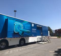
In June, the Philips Radiology Experience Tour hit the road to provide healthcare professionals with an opportunity to ...
September 19, 2023 
News | X-Ray
April 29, 2026 — Results from a new study* presented at the American Roentgen Ray Society’s (ARRS) 2026 annual meeting ...
April 29, 2026 
News | Imaging Software Development
April 28, 2026 — Avatar Medical has been granted FDA 510(k) clearance for Avatar Medical Vision, its software platform ...
April 28, 2026 News | Cardiac Imaging
April 28, 2026 — Abbott has received U.S. Food and Drug Administration (FDA) clearance and CE Mark for its next ...
April 28, 2026 Sponsored Content
Videos | Radiology Imaging
This summer, the Philips Radiology Experience Tour has been bringing Philips imaging modalities directly to the ...
August 14, 2023 
News | FDA
April 16, 2026 — Royal Philips has received U.S. Food and Drug Administration 510(k) clearance for the Philips Spectral ...
April 20, 2026 
News | Radiology Imaging
April 7, 2026 — Onvida Health and Siemens Healthineers have entered a 10-year Value Partnership¹ designed to bring the ...
April 09, 2026 
News | Computed Tomography (CT)
April 2, 2026 — Nano-X Imaging Ltd. recently announced its U.S.-based subsidiary, Nanox Impact Inc., has signed a new ...
April 08, 2026 Sponsored Content

Case Study | Enterprise Imaging
The healthcare industry faces many different types of obstacles in today’s challenging marketplace. Staff shortages ...
August 01, 2023  Clearance to GE HealthCare's True Definition DL Software")
News | Computed Tomography (CT)
April 2, 2026 — GE HealthCare has received 510(k) clearance from the U.S. Food and Drug Administration (FDA) for True ...
April 03, 2026 
News | Computed Tomography (CT)
March 30, 2026 — HCA Healthcare’s Good Samaritan Hospital is the first hospital in the Bay Area to implement Lumina 3D ...
April 01, 2026 
News | Radiology Business
March 31, 2026 — Radon Medical Imaging, a medical imaging equipment maintenance and repair services company, has has ...
March 31, 2026 
News | Radiology Imaging
March 26, 2026 — GE HealthCare has announced a renewed research collaboration with Stanford Medicine Department of ...
March 30, 2026 
News | Cardiac Imaging
March 28, 2026 — When Ashley Perlow felt a sharp pain shoot across her chest and into both wrists, she didn't think it ...
March 30, 2026 
News | FDA
March 24, 2026 — MARS Bioimaging, a New Zealand–headquartered medical device company, has received U.S. Food and Drug ...
March 25, 2026 
News | Computed Tomography (CT)
March 23, 2026 — GE HealthCare has received 510(k) clearance from the U.S. Food and Drug Administration (FDA) for ...
March 23, 2026 
News | Radiology Imaging
March 23, 2026 — Samsung Medison hsa announced that its U.S. medical imaging businesses, previously operating as ...
March 23, 2026 © Copyright Wainscot Media. All Rights Reserved.
Subscribe Now