
May 24, 2024 — Smokers who have small abnormalities on their CT scans that grow over time have a greater likelihood of ...
Computed Tomography (CT)
Computed tomography (CT) systems use a series of X-ray images to create an image volume dataset with slices that can be manipulated on any plane using advanced visualization software. The section includes computed tomography scanners, CT contrast agents, CT angiography (CTA and CCTA), CT perfusion, spectral CT (dual-source CT), and iterative reconstruction dose reduction software.
News | Radiology Business
May 22, 2024 — Medtronic has announced new preliminary data from the VERITAS clinical study using its ILLUMISITE ...
 May 22, 2024
News | Artificial Intelligence
May 15, 2024 — Heart disease is the leading cause of mortality in the U.S., accounting for one out of every five deaths ...
May 15, 2024 Sponsored Content

News | Computed Tomography (CT)
SPONSORED CONTENT — Fujifilm’s latest CT technology brings exceptional image quality to a compact and user- and patient ...
August 06, 2024 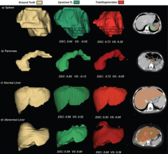
News | Pediatric Imaging
May 15, 2024 — Transfer learning (TL) models trained on heterogeneous public datasets and fine-tuned using institutional ...
May 15, 2024 
News | Radiology Business
May 14, 2024 — University Hospitals (UH) and Siemens Healthineers announce a 10-year strategic alliance that builds on ...
May 14, 2024 
News | Treatment Planning
May 6, 2024 — Elekta announced the acquisition of Philips Healthcare’s Pinnacle Treatment Planning System (TPS) patent ...
May 06, 2024 Sponsored Content

Case Study | Radiology Imaging
In June, the Philips Radiology Experience Tour hit the road to provide healthcare professionals with an opportunity to ...
September 19, 2023 
News | Pediatric Imaging
May 2, 2024 — Head and abdominal trauma is a leading cause of death for children. About 1%–2% of children who come to ...
May 02, 2024 
Feature | Radiology Business
Beginning this spring, ITN will begin sending out a bi-monthly survey to our readers on a variety of topics, which we ...
May 02, 2024 
News | Enterprise Imaging
April 25, 2024 — International medical imaging IT and cybersecurity company Sectra has signed two contracts to provide ...
April 25, 2024 Sponsored Content
Videos | Radiology Imaging
This summer, the Philips Radiology Experience Tour has been bringing Philips imaging modalities directly to the ...
August 14, 2023 
News | Radiology Business
April 23, 2024 — A diverse writing group, led by authors at the University of Toronto, have developed an approach for ...
April 23, 2024 
News | Computed Tomography (CT)
April 22, 2024 — A new study showed that a non-invasive imaging test can help identify patients with coronary artery ...
April 22, 2024 
News | Clinical Trials
April 16, 2024 — QT Imaging Holdings, Inc., a medical device company engaged in research, development, and ...
April 16, 2024 Sponsored Content

Case Study | Enterprise Imaging
The healthcare industry faces many different types of obstacles in today’s challenging marketplace. Staff shortages ...
August 01, 2023 
News | Mammography
April 12, 2024 — Bayer and Hologic, Inc. announced a first-of-its-kind collaboration to deliver a coordinated solution ...
April 12, 2024 
News | Mammography
April 12, 2024 — GE HealthCare, a leader in breast health technology and diagnostics, will feature its latest breast ...
April 12, 2024 
News | Radiation Therapy
March 28, 2024 — RefleXion Medical, Inc., a therapeutic oncology company, and Limbus AI, Inc., a provider of software ...
March 28, 2024 
News | Artificial Intelligence
March 18, 2024 — RamSoft, a global leader in novel cloud-based RIS/PACS radiology solutions for imaging centers and ...
March 18, 2024 
News | Breast Imaging
March 18, 2024 — QT Imaging Holdings, Inc., a medical device company engaged in research, development, and ...
March 18, 2024 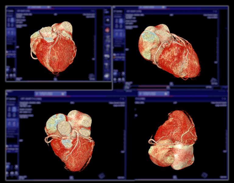
Feature | Computed Tomography (CT) | By Melinda Taschetta-Millane
Computed Tomography (CT) continues to be a rapidly evolving technology with many new advancements, as displayed and ...
March 07, 2024 
News | Radiology Imaging
March 5, 2024 — Life Guard Imaging, a pioneering leader in preventative imaging services, is thrilled to announce its ...
March 05, 2024 
News | Artificial Intelligence
February 29, 2024 — AIxSCAN, Inc., a Sunnyvale, CA-based developer of a next generation artificial intelligence (AI) ...
February 29, 2024 © Copyright Wainscot Media. All Rights Reserved.
Subscribe Now