
October 3, 2023 — Using previously taken diagnostic computed tomography (CT) scans in place of CT simulation scans to ...
Computed Tomography (CT)
Computed tomography (CT) systems use a series of X-ray images to create an image volume dataset with slices that can be manipulated on any plane using advanced visualization software. The section includes computed tomography scanners, CT contrast agents, CT angiography (CTA and CCTA), CT perfusion, spectral CT (dual-source CT), and iterative reconstruction dose reduction software.

News | Computed Tomography (CT)
September 29, 2023 —Nano-X Imaging, an innovative medical imaging technology company, today announced that HealthCCSng ...
 September 29, 2023
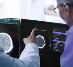
News | Artificial Intelligence
September 20, 2023 — Medical imaging artificial intelligence (AI) company Annalise.ai has announced that the results ...
September 20, 2023 Sponsored Content

News | Computed Tomography (CT)
SPONSORED CONTENT — Fujifilm’s latest CT technology brings exceptional image quality to a compact and user- and patient ...
August 06, 2024 
Sponsored Content | Case Study | Radiology Imaging | By Tim Hodson
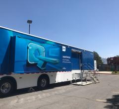
In June, the Philips Radiology Experience Tour hit the road to provide healthcare professionals with an opportunity to ...
September 19, 2023 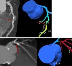
News | Computed Tomography (CT)
September 19, 2023 — An advanced CT test can identify individuals with stable angina at a reduced risk of three-year ...
September 19, 2023 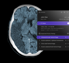
News | FDA
September 13, 2023 — Annalise.ai, a leader in AI-powered medical imaging solutions, announced the receipt of 510(k) ...
September 13, 2023 Sponsored Content
Case Study | Radiology Imaging
In June, the Philips Radiology Experience Tour hit the road to provide healthcare professionals with an opportunity to ...
September 19, 2023 
September 8, 2023 — A simple, noninvasive contrast enhanced ultrasound (CEUS) scan is an ideal tool for resolving ...
September 08, 2023 
Feature | PET Imaging
According to a new report from Transparency Market Research (TMR), PET radiotracers are expected to rise at a CAGR of 8 ...
September 05, 2023 
News | Radiology Imaging
August 24, 2023 — Medical imaging via X-rays, CT scans, MRIs and ultrasounds provide health-care professionals with ...
August 24, 2023 Sponsored Content
Videos | Radiology Imaging
This summer, the Philips Radiology Experience Tour has been bringing Philips imaging modalities directly to the ...
August 14, 2023 
News | Radiology Business
August 17, 2023 — SSM Health and Siemens Healthineers have announced a new 10-year strategic partnership agreement ...
August 17, 2023 
News | Contrast Media
August 17, 2023 — University of Missouri School of Medicine neurologist Adnan Qureshi, MD recently led a study that ...
August 17, 2023 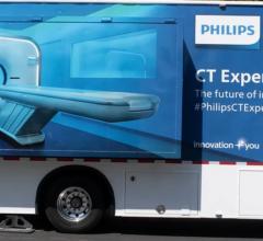
Sponsored Content | Videos | Radiology Imaging
This summer, the Philips Radiology Experience Tour has been bringing Philips imaging modalities directly to the ...
August 14, 2023 Sponsored Content

Case Study | Enterprise Imaging
The healthcare industry faces many different types of obstacles in today’s challenging marketplace. Staff shortages ...
August 01, 2023 
News | Computed Tomography (CT)
August 14, 2023 — Precision Imaging Centers has added Fujifilm Healthcare Americas Corporation’s Scenaria View low-dose ...
August 14, 2023 
August 14, 2023 — GigXR, Inc, whose immersive learning platform delivers a catalog of complementary XR applications for ...
August 14, 2023 
News | Artificial Intelligence
August 9, 2023 — Annalise.ai, one of the global leaders of AI decision-support solutions, announced that their Annalise ...
August 09, 2023 
News | Artificial Intelligence
August 7, 2023 — AI tools that quickly and accurately create detailed narrative reports of a patient’s CT scan or X-ray ...
August 07, 2023 
News | Artificial Intelligence
August 7, 2023 — GigXR, Inc, whose immersive learning platform delivers a catalog of complementary XR applications for ...
August 07, 2023 
Sponsored Content | Case Study | Enterprise Imaging
The healthcare industry faces many different types of obstacles in today’s challenging marketplace. Staff shortages ...
August 01, 2023 
News | Computed Tomography (CT)
July 31, 2023 — The American College of Radiology (ACR) has issued a statement regarding ACP colorectal cancer screening ...
July 31, 2023 
News | AHRA
July 24, 2023 — The Association for Medical Imaging Management (AHRA) announced that GE Healthcare won the 2023 ...
July 24, 2023 © Copyright Wainscot Media. All Rights Reserved.
Subscribe Now