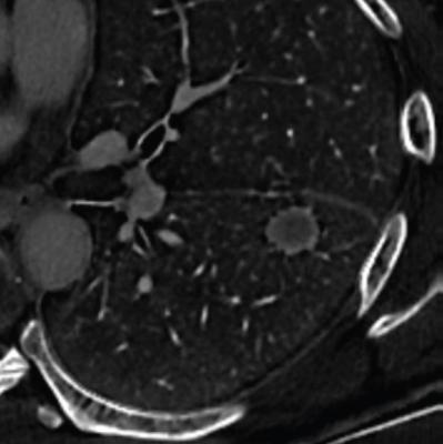
62-year-old man with history of oropharyngeal cancer, treated by chemoradiation therapy. Contrast-enhanced multiphase DECT of chest performed for surgical planning. Axial image from iodine concentration map, reconstructed from equilibrium-phase acquisition, shows thin ringlike peripheral high iodine concentration, involving nodule’s entire circumference. Nodule underwent resection, revealing oropharyngeal carcinoma metastasis. Case represents true-positive example of using ringlike peripheral high iodine concentration for diagnosis of pulmonary metastasis.
January 11, 2023 — According to an accepted manuscript published in ARRS’ American Journal of Roentgenology (AJR), ringlike peripheral high iodine concentration maps from dual-energy CT (DECT) can help guide management in patients with known lung cancer and an indeterminate solitary nodule.
“Ringlike peripheral high iodine concentration had excellent interobserver agreement, showed high specificity (albeit poor sensitivity) for differentiating pulmonary metastasis from primary lung cancer, and independently predicted pulmonary metastasis,” wrote AJR first author Yoshinao Sato, MD, PhD, from the Diagnostic Imaging Center at Japan’s Cancer Institute Hospital in Tokyo.
This AJR accepted manuscript study included 93 patients (64 men, 29 women; median age, 70 years) who underwent resection of a primary lung cancer (n=68) or pulmonary metastasis (n=25) corresponding with a solid lesion on preoperative contrast-enhanced DECT performed between April 2020 and March 2021. After constructing venous-phase 120-keV single-energy images, equilibrium-phase 66-keV virtual monoenergetic images, as well as iodine concentration maps, two radiologists independently assessed lesions for the following: spiculated margins, air bronchograms, rim enhancement, and thin ringlike peripheral high iodine concentration.
Ultimately, ringlike peripheral high iodine concentration on DECT showed excellent interobserver agreement (κ=0.80), and had sensitivity of 52% and specificity of 81% for differentiating pulmonary metastases from primary lung cancers. Additionally, the finding independently predicted pulmonary metastasis in multivariate analysis [OR=7.81, 95% CI: 2.28–29.60; p=.001] combining patient and lesion characteristics.
“Iodine concentration maps from DECT could help determine the diagnosis for lesions that are equivocal for pulmonary metastasis on conventional images,” the authors of this AJR accepted manuscript reiterated.
For more information: www.arrs.org

 July 15, 2026


