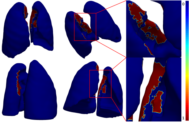
A new study led by researchers at Emory AI.Health, published in the Journal of Computers in Medicine and Biology, sheds light on the significant lung damage caused by severe COVID-19. In this image, AI highlights areas of the lungs with architectural distortions. Blue areas indicate mild COVID-19, while red areas indicate severe COVID-19. Image courtesy: Emory University
June 11, 2024 — A new study led by researchers at Emory AI.Health, published in the Journal of Computers in Medicine and Biology, has shed light on the significant lung damage caused by severe COVID-19, according to the study authors, who emphasized that as the immediate threat of COVID-19 subsides, investigating the virus’s lasting impacts remains essential.
“Although the acute phase of COVID-19 has largely been mitigated, the persistence and impact of long COVID remains a concern. Our AI analysis identified specific areas of lung damage that could have enduring consequences,” said Anant Madabhushi, PhD, Executive Director of Emory AI.Health and principal investigator of the study. “While we have not yet examined long COVID patients explicitly, it’s crucial to investigate whether these individuals exhibit residual lung deformation, which could provide valuable insights into the long-term effects of this disease,” he added.
About the AI Lung Study
The study, “Deep learning reveals lung shape differences on baseline chest CT between mild and severe COVID-19: A multi-state retrospective study,” utilized artificial intelligence (AI) to analyze CT scans from over 3,400 patients across multiple institutions. These patients were categorized into three groups: healthy individuals, those with mild COVID-19, and those with severe COVID-19 requiring ventilator support.
The analysis revealed that patients with severe COVID-19 experienced significant deformities to the surfaces of the lungs, particularly along the mediastinal and basal surfaces. These findings are critical for understanding the potential long-term effects of the disease. Researchers suggest that these lung deformations are likely to impair lung function, ultimately affecting human health and quality of life, and potentially increasing overall mortality.
The role of AI in lung analysis
The AI technology used in the study employed a 3D residual convolutional network to segment and analyze lung images, according to a written summary of the study issued by Emory University. This approach, it added, allowed researchers to create a detailed map of lung deformations, identifying specific areas of damage. By comparing lung images of healthy individuals with those of mild and severe COVID-19 patients, the AI provided a comprehensive map of lung deformations, highlighting the extent of the damage caused by severe COVID-19.
One of the study's goals was to demonstrate how AI can systematically evaluate the long-term effects of COVID-19 on internal organs. The ability to identify and quantify lung damage through AI not only helps researchers understand the immediate impact of severe COVID-19, but also offers insight into the potential lingering effects on lung function.
“COVID-19 can cause serious complications such as pneumonia, severe lung damage, and blood infections, which can lead to lasting lung problems like scarring and chronic breathing issues,” said Amogh Hiremath, AI scientist at Picture Health and first author of the study. “While some people recover fully, others may suffer permanent lung damage. Understanding how COVID-19 affects the lungs during its early onset can help us better understand and treat the disease,” added Hiremath.
More information: www.emory.edu, www.aihealth.emory.edu
Reference: DOI: 10.1016/j.compbiomed.2024.108643

 October 07, 2026