
Dec. 12. 2025 — A new study has found that an individualized approach to breast cancer screening that assesses patients’ ...
Breast Imaging
Women's health related to breast imaging, including mammography, breast MRI, ABUS, automated breast ultrasound, breast ultrasound, breast biopsy, PEM and positron emission mammography.

News | Breast Imaging
Dec. 16, 2025 — Hologic, Inc, a medical technology company dedicated to improving women’s health, recently announced new ...
 December 16, 2025
News | Artificial Intelligence
Dec. 1, 2025 — Researchers at the University of California, Berkeley and University of California, San Francisco have ...
December 10, 2025 Sponsored Content

Case Study | Artificial Intelligence
As the largest independent imaging group in Michigan with 10 locations across the state, Regional Medical Imaging (RMI) ...
April 09, 2020 
News | Breast Imaging
Dec. 01, 2025 — DeepHealth, a wholly owned subsidiary of RadNet, Inc., has launched the DeepHealth Breast Suite,2 an end ...
December 04, 2025 
News | Women's Health
Dec. 1, 2025 — ScreenPoint Medical has completed a commercial agreement making its Transpara breast-imaging AI portfolio ...
December 03, 2025 
News | Mammography
Nov. 30, 2025 — At RSNA 2025, Siemens Healthineers will introduce new capabilities for its Mammomat B.brilliant ...
December 02, 2025 Sponsored Content
Case Study | Breast Imaging
Christina Jacobs, M.D., Director of Breast Imaging (Bronson Health System) is always looking for ways to work more ...
April 09, 2020 
News | RSNA 2025
Dec. 2, 2025 — Lunit, a provider of AI for cancer diagnostics and precision oncology, will present 14 studies at RSNA ...
December 02, 2025 
News | Women's Health
Dec. 1, 2025 — A study of data from seven outpatient facilities in the New York region found that 20-24% of all the ...
December 02, 2025 
News | Artificial Intelligence
Nov. 25, 2025 – Medical imaging AI company Avicenna.AI has announced a strategic partnership with Ferrum, an AI ...
November 25, 2025 Sponsored Content
Videos | Artificial Intelligence
ProFound AI is an FDA-cleared artificial intelligence (AI) system for reading 3-D breast tomosynthesis images. At RSNA19 ...
February 06, 2020 
News | Ultrasound Imaging
Nov. 12, 2025 — GE HealthCare and DeepHealth, Inc., a wholly owned subsidiary of RadNet, Inc., have announced their ...
November 20, 2025 
News | Breast Imaging
Nov. 17, 2025 — RadNet, Inc. and its wholly owned subsidiary, DeepHealth have announced results from the largest real ...
November 17, 2025 
News | Artificial Intelligence
Nov. 6, 2025 — Lunit, a provider of AI for cancer diagnostics and precision oncology, recently announced that Volpara ...
November 07, 2025 Sponsored Content
Videos | Digital Radiography (DR)
At RSNA19, David Widmann, president and CEO of Konica Minolta Healthcare Americas, discussed innovation, stressing the ...
January 03, 2020 
News | RSNA 2025
Nov. 3, 2025 — QT Imaging Holdings has announced that its chief medical officer, Elaine luanow, MD, will host a seminar ...
November 04, 2025 
News | Women's Health
Nov. 3, 2025 — —A new radioimmunotherapy approach has the potential to cure human epidermal growth factor receptor 2 ...
November 04, 2025 
News | Breast Imaging
Oct. 28, 2025 — QT Imaging Holdings, Inc., a medical device company focused on radiation-free imaging technology, has ...
October 28, 2025 
Sponsored Content | Feature | Breast Imaging
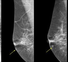
Despite decades of progress in breast imaging, one challenge continues to test even the most skilled radiologists ...
October 24, 2025 
News | Breast Imaging
Oct. 15, 2025 — Leading into Breast Cancer Awareness Month, Fujifilm Healthcare Americas Corp. and Beekley Medical ...
October 15, 2025 
News | RSNA 2025
Oct. 7, 2025 – Clairity Inc., a leader in AI-based breast cancer risk prediction, will make five scientific ...
October 07, 2025 
News | Breast Imaging
Oct. 3, 2025 — Gnosis for Her, a mobile breast health initiative redefining comfort and access in women's breast imaging ...
October 06, 2025 
News | Mammography | Mayo Clinic
Early detection is key to breast cancer survival. But nearly half of all women in the U.S. have dense breast tissue ...
October 03, 2025 © Copyright Wainscot Media. All Rights Reserved.
Subscribe Now