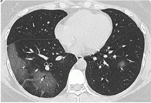
Series CT scans in 35-year-old woman with COVID-19 pneumonia. (a) Scan obtained on illness days 1 showed multiple pure ground-glass opacity (GGO) mainly in right lower lobe. (b) Scan obtained on illness days 5 showed increased extent of GGO and early consolidation. (c) Scan obtained on illness days 11 showed multiple consolidation with almost the same extent. (d) Scan obtained on illness days 15 showed a mixed pattern with a slightly smaller extent, and the perilobular consolidation might suggest the presence of organizing pneumonia. The patient was discharged on illness days 17. Image courtesy of the journal Radiology
March 20, 2020 — In a new study published in the journal Radiology, researchers found mild to significant lung abnormalities on the chest computed tomography (CT) images of 94 percent of patients with COVID-19 upon discharge from the hospital, suggesting follow-up monitoring of patients might be necessary.
The researchers performed a longitudinal study to analyze the serial CT findings over time in patients with COVID-19. During Jan. 16 to Feb. 17, 2020, 90 patients (33 men and 57 women, mean age 45 years) with COVID-19 pneumonia were prospectively enrolled and followed up through discharge, death or study end. Radiologists reviewed 366 CT scans for the patterns and distribution of lung abnormalities, total CT scores and number of zones involved. Those features were analyzed for temporal change.
The extent of CT abnormalities progressed rapidly after symptom onset and peaked during illness days 6-11. The predominant pattern of abnormalities after symptom onset was ground-glass opacity. Sixty-six of the 70 patients discharged had residual disease on final CT scans, with ground-glass opacity the most common pattern.
“A better understanding of the progression of CT findings in COVID-19 pneumonia may help facilitate accurate diagnosis and disease stage,” the authors wrote.
You can read the full study: Temporal Changes of CT Findings in 90 Patients with COVID-19 Pneumonia: A Longitudinal Study.
Find the latest Radiology and Radiology: Cardiothoracic Imaging COVID-19 research at Special Focus: COVID-19.
Related Coronavirus Content:
VIDEO: Imaging COVID-19 With Point-of-Care Ultrasound (POCUS)
The Cardiac Implications of Novel Coronavirus
CT Provides Best Diagnosis for Novel Coronavirus (COVID-19)
Radiology Lessons for Coronavirus From the SARS and MERS Epidemics
Deployment of Health IT in China’s Fight Against the COVID-19 Epidemic
Emerging Technologies Proving Value in Chinese Coronavirus Fight
Radiologists Describe Coronavirus CT Imaging Features
Coronavirus Update from the FDA
CT Imaging of the 2019 Novel Coronavirus (2019-nCoV) Pneumonia
CT Imaging Features of 2019 Novel Coronavirus (2019-nCoV)
Chest CT Findings of Patients Infected With Novel Coronavirus 2019-nCoV Pneumonia
Find more related clinical content Coronavirus (COVID-19)
ACC COVID-19 recommendations for the cardiovascular care team
VIDEO: What Cardiologists Need to Know about COVID-19 — Interview with Thomas Maddox, M.D.
The Cardiac Implications of Novel Coronavirus
ESC Council on Hypertension Says ACE-I and ARBs Do Not Increase COVID-19 Mortality
Additional COVID-19 Resources for Clinicians:
Johns Hopkins Coronavirus Resource Center with inteavtive map of cases in U.S. and worldwide
World Health Organization (WHO) COVID-19 situation reports
World Health Organization (WHO) coronavirus information page
U.S. Food and Drug Administration (FDA) COVID-19 information page

 July 20, 2026


