#COVID19 #Coronavirus #2019nCoV #Wuhanvirus #SARScov2

Getty images


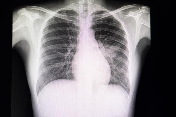
April 14, 2020 — Chest X-rays are often used to detect infections in the lungs, but the world’s largest study of its kind finds it’s not a reliable way to diagnose respiratory infections caused by COVID-19

The research team led by Michael Weinstock, M.D., an adjunct professor of emergency medicine at The Ohio State University College of Medicine, reviewed more than 630 chest X-rays of confirmed and symptomatic COVID-19 patients of a large urgent care company in New York and New Jersey. The radiologists determined the chest x-rays were normal in 58.3% of cases, and normal or only mildly abnormal in 89% of patients.

“Providers ordering a chest x-ray in the outpatient setting should be aware that a patient with symptoms of COVID-19 may have a negative chest X-ray and should manage the patient based on their symptoms,” said Weinstock. “Doctor’s should not be reassured by a negative chest X-ray.”

Additionally, the research in The Journal of Urgent Care Medicine found the chest x-ray results of COVID-19 patients differ from those of bacterial pneumonia patients. The abnormal chest x-ray findings of COVID-19 patients were more likely to be scattered diffusely throughout both of the lungs than typical pneumonia.

“This study reinforces what we’ve learned from our colleagues outside of the United States that the majority of patients with COVID-19 have mild symptoms and minimal evidence of disease on chest X-ray,” said Matthew Exline, M.D., associate professor in the department of Pulmonary, Critical Care and Sleep Medicine at The Ohio State University College of Medicine and medical director of the medical intensive care unit at The Ohio State University Wexner Medical Center. “Hopefully this will help clinicians decide who would best benefit from hospitalization and any potential new treatments.”

Researchers are working on a second study to look at the outcomes of these patients and how it correlates to their chest X-ray results.


Related Content

News | Artificial Intelligence

September 25, 2023 — Viz.ai, a leader in AI-powered disease detection and intelligent care coordination, today announced ...

Time September 25, 2023
arrow
Feature | Magnetic Resonance Imaging (MRI) | Robert L. Bard, MD, and Lennard M. Gettz, EdD

Since the advent of the magnetic resonance imaging (MRI) exam on human patients in the late 1970s, this innovation ...

Time September 25, 2023
arrow
News | Cardiac Imaging

September 21, 2023 — Declines in cardiovascular procedure volumes observed early in the COVID-19 pandemic greatly ...

Time September 21, 2023
arrow
News | X-Ray

September 21, 2023 — Scientists in Moscow have successfully engineered a prototype detector for X-ray and PET/CT ...

Time September 21, 2023
arrow
News | Enterprise Imaging

September 20, 2023 — aycan, a recognized leader in medical imaging, announced today a new solution for reviewing DICOM ...

Time September 20, 2023
arrow
Feature | Radiation Dose Management | By Melinda Taschetta-Millane

Early-stage detection is key to the prevention of life-threatening diseases; however, limited access to data hinders the ...

Time September 20, 2023
arrow
Feature | Ultrasound Imaging | By Mustafa Hassan, PhD

Handheld ultrasound has not yet reached mainstream adoption, but the market is still forecast to reach over $500 million ...

Time September 20, 2023
arrow
News | Radiology Business

September 20, 2023 — GE HealthCare released its inaugural Sustainability Report, which describes the steps the company ...

Time September 20, 2023
arrow
Sponsored Content | Case Study | Radiology Imaging | By Tim Hodson

In June, the Philips Radiology Experience Tour hit the road to provide healthcare professionals with an opportunity to ...

Time September 19, 2023
arrow
Feature | Magnetic Resonance Imaging (MRI) | By Christine Book

The world of medical imaging is marking a significant milestone in 2023: the 50th anniversary of magnetic resonance ...

Time September 19, 2023
arrow
Subscribe Now