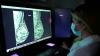
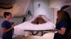
Constance "Connie" Lehman, M.D., Ph.D., chief of breast imaging, co-director of the Avon Comprehensive Breast Evaluation Center at the Massachusetts General Hospital, and professor of radiology at Harvard Medical School, explains issues and suggested guidelines for women who receive the COVID-19 vaccine and need to get a mammogram. In the first three months since the vaccines have been released, there have been numerous case reports of the vaccine causing swollen lymph nodes. This is would usually raise a red flag for breast cancer, but is normal for many women receiving the vaccine as their body's immune system gears up against the virus.
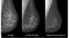
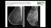
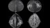
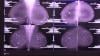
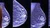
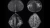
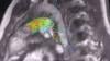
Lehman said cases reports of axillary adenopathy have been identified on breast imaging after coronavirus disease (COVID-19) vaccination and are rising. Lehman et al. proposed a pragmatic management approach in a recent article in the American Journal of Roentgenology (AJR).[1]
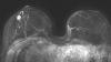
In the settings of screening mammography, screening MRI and diagnostic imaging work-up of breast symptoms, with no imaging findings beyond unilateral axillary adenopathy ipsilateral to recent (prior six weeks) vaccination, they report the adenopathy as benign with no further imaging indicated if no nodes are palpable six weeks after the last vaccine dose.
For patients with palpable axillary adenopathy in the setting of ipsilateral recent vaccination, clinical follow-up of the axilla is recommended. In all these scenarios, axillary ultrasound is recommended if clinical concern persists six weeks after vaccination.
In patients with recent breast cancer diagnosis in the pre- or peri-treatment setting, prompt recommended imaging is encouraged as well as vaccination (in the thigh or contralateral arm). The recommendations align with the ACR BI-RADS Atlas and aim to: 1) reduce patient anxiety, provider burden, and costs of unnecessary evaluation of enlarged nodes in the setting of recent vaccination, and 2) avoid further delays in vaccinations and breast cancer screening during the pandemic.
Related Medical Imaging of COVID Content:
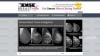
COVID-19 Vaccination Axillary Adenopathy Detected During Breast Imaging
CMS Now Requires COVID-19 Vaccinations for Healthcare Workers by January 4
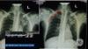
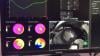
PHOTO GALLERY: How COVID-19 Appears on Medical Imaging
VIDEO: Imaging COVID-19 With Point-of-Care Ultrasound (POCUS) — Interview with Mike Stone, M.D.
VIDEO: Use of Teleradiology During the COVID-19 Pandemic — Interview with John Kim, M.D.
Find more radiology related COVID content
Reference:
1. Constance D. Lehman, Leslie R. Lamb, and Helen Anne D'Alessandro. Mitigating the Impact of Coronavirus Disease (COVID-19) Vaccinations on Patients Undergoing Breast Imaging Examinations: A Pragmatic Approach American Journal of Roentgenology. 10.2214/AJR.21.25688