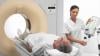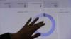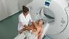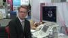
At RSNA 2016, the key buzzwords were “deep learning,” “machine learning” and “artificial intelligence.” Vendors and major academic centers are developing a wide array of artificial intelligence neural networks to aid radiologists in clinical diagnosis and clinical decision support. In the future, AI may also be able to help train radiologists on both normal and abnormal presentations of various organs and body systems so as to more easily identify related disease states and conditions. The following video offers two examples of how the IBM Watson system examines imaging studies.
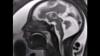
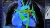
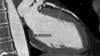
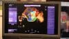
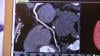
The first case seen here demonstrates how Watson can arrive at a differential diagnosis of an aortic dissection by analyzing an abdominal computed tomography (CT) scan. The second case involves the discovery of a fibroadenoma of the breast from Watson’s analysis of a mammogram.
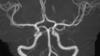
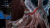
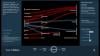
Watson first analyzes the text of the radiology report, identifying and pulling out key words or phrases that may indicate the potential diagnosis. It then examines the CT scan to locate relevant visible anatomic structures such as the heart, aorta and pulmonary artery. Each structure is examined for anomalies, which identifies a possible aortic dissection; the dissection is displayed within the context of the entire 3-D CT scan. Finally, Watson applies its existing clinical knowledge to the findings from the CT scan and the radiology report, establishing pathways to numerous possible conclusions until arriving at the right one.
See examples of real products using AI at RSNA 2017 in the VIDEO "Examples of How Artificial Intelligence Will Improve Medical Imaging." ITN also created an in-depth VIDEO: Technology Report — Artificial Intelligence at RSNA 2017, with interviews with numerous AI vendors.
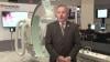
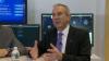
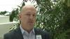
Watch the VIDEO: “Development of Artificial Intelligence to Aid Radiology,” an interview with Mark Michalski, M.D., director of the Center for Clinical Data Science at Massachusetts General Hospital, explaining the basis of artificial intelligence in radiology.