The Institute of Medicine (IOM) of the National Academies issued its report, “Delivering High-Quality Cancer Care: Charting a News Course for a System in Crisis.” The 315-page report, produced by a 17-member committee of cancer care leaders, concludes that the nation’s “cancer care delivery system is in crisis. Care is not patient-centered, many patients do not receive palliative care to manage their symptoms and side effects from treatment, and decisions about care often are not based on the latest scientific evidence.” In addition, the report details a six-part framework for improving the quality of cancer care, improving “the quality of life and outcomes for people facing a cancer diagnosis.”
At RSNA 2013, Carestream’s new Vue PACS (picture archive and communication system) Reporting (shown as works-in-progress) is designed to enable insertion of key images and quantitative comparisons such as vessel analysis and measurements from modalities into radiology reports, which is designed to aid clinicians in treatment planning.
The QA modules of Standard Imaging Inc. PIPSpro feature a new, simplified user-interface for TG-142 testing. Since the ...
Radiology departments have many different needs and face a wide variety of challenges that can impact their departments ...
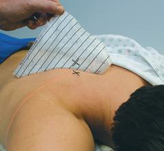
A light, medical grade, all-over adhesive ensures Beekley Medical’s new Mark-Through GuideLines stay in place without buckling or lifting during computed tomography (CT)-guided biopsies. Marks can be made anywhere on grid. The ink instantly transfers through to clearly marked entry sites on patient’s skin. Evenly spaced, lead-free radiopaque lines provide a distinct artifact-free image further enhancing the accuracy of needle placement and mark correlation.
At RSNA 2013, Toshiba will introduce a new single-panel Radrex-i wireless digital radiographic (DR) system X-ray system as a works-in-progress. The single panel wireless solution lowers cost of ownership while enabling more out-of-Bucky work and increasing workflow with one of the lightest detectors on the market.
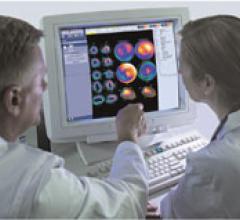
UltraSPECT announced that MaineHealth, a family of hospitals and medical centers throughout Maine, has selected the UltraSPECT Xpress.Cardiac and Xpress3.Cardiac solutions as part of the organization’s strategy for reducing nuclear medicine (NM) dose and complying with the American Society of Nuclear Cardiology (ASNC) final guidelines effective Jan. 1, 2014. Maine Medical Partners (MMP) – MaineHealth Cardiology in South Portland is one of ten facilities within MaineHealth that is installing the product, with others scheduled to continue roll out before the end of 2013.
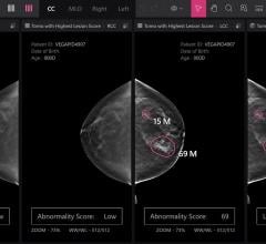
Despite decades of progress in breast imaging, one challenge continues to test even the most skilled radiologists ...
Cancer patients from all over the world now have a new opportunity to access proton therapy today – at the Proton Therapy Center Czech in Prague. It is only the fifth such center in Europe and currently the most well equipped centre in the world. The facility is attracting child and adult cancer patients from around the world seeking advanced cancer care with few treatment-related side effects.
In patients with atrial fibrillation, delayed enhancement magnetic resonance imaging (DE-MRI) performed before ablative treatment can stage the degree of damaged heart tissue (atrial fibrosis) and help predict whether treatment will be successful or not, according to results of Delayed Enhancement — MRI determinant of successful Catheter Ablation of Atrial Fibrillation (DECAAF) trial.
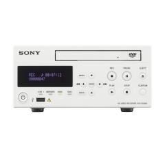
At RSNA 2013, Sony will be showing a works-in-progress medical recorder, the HVO-500MD (without DVD optical drive) and the HVO-550MD (with DVD optical drive). These models are designed specifically for medical recording.
Bayer Radiology’s Barbara Ruhland and Thom Kinst discuss how radiology departments can address the many different ...
Toshiba is introducing a new Aquilion One platform at RSNA 2013. The new Aquilion One family allows providers to more precisely choose the system that fits their needs today while giving them an upgrade path for the future.
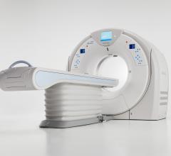
September 9, 2013 — Toshiba’s new Aquilion Prime is a versatile, scalable and patient-friendly computed tomography (CT) system. The system’s technologies are built for the clinical and financial needs of users both in the present and future.
The U.S. Food and Drug Administration (FDA) has granted 510(k) market clearance for Siemens’ Symbia Intevo, the first system to completely integrate high sensitivity of single-photon emission computed tomography (SPECT) with the high specificity of computed tomography (CT) into a single modality, rather than imaging as separate overlay images.
eHealth Saskatchewan plays a vital role in providing IT services to patients, health care providers, and partners such ...
At the 2013 Radiological Society of North America’s annual meeting (RSNA 2013), aycan, a provider of cost-saving PACS solutions, will highlight new release plugins for its post-processing workstation, aycan OsiriX PRO, which significantly expands the functionality of its specialty workstations for mammography, oncology and vascular surgery.
While the advantages of digitalizing a hospital can be multiplied across a multi-site enterprise, the challenges can be magnified as well. That’s why Chirec, with its six sites, in Brussels, Belgium, has a comprehensive strategy to go digital and become "paperless." Agfa HealthCare’s ICIS View is bringing the hospital’s images and users closer together, and Chirec one step closer to a completely digital future.
A Memorial Sloan-Kettering Cancer Center study shows that the use of magnetic resonance imaging (MRI) before or immediately after surgery in women with ductal carcinoma in situ (DCIS) was not associated with reduced local recurrence or contralateral breast cancer rates.
More than two decades after becoming the world’s first hospital-based proton therapy center, the engineers, scientists and physicians at Loma Linda University Medical Center (LLUMC) continue to develop new technologies that will lead to the next generation of proton therapy treatment.
Reportlinker.com released a new report on the MRI market titled “Advances in Magnetic Resonance Imaging (MRI) Market (2013-2018) - Technology Trend Analysis – By Architecture, Field Strengths, Technology & Applications in Medical Diagnostics with Market Landscape Analysis – Estimates up to 2018.”
For patients with degenerative cervical myelopathy, imaging with 18F-FDG positron emission tomography (PET) could act as a marker for a potentially reversible phase of the disease in which substantial clinical improvement can be achieved. According to research published in the September issue of The Journal of Nuclear Medicine, patients who exhibited hypermetabolism at the point of compression in their spine experienced improved outcomes after undergoing decompressive surgery.
American Shared Hospital Services announced today that the Centers for Medicare and Medicaid Services (CMS) has posted a revision of the previously proposed rule for Medicare's hospital outpatient prospective payment system for calendar year 2014. Within this proposed rule, CMS proposes updates for the delivery codes used for stereotactic radiosurgery (SRS), including Gamma Knife services.
For over 70 years, Insight Medical Imaging has provided state-of-the-art medical imaging services to the city of Edmonton and its surrounding areas. Having started out as a smaller, hospital-based imaging organization, Insight has grown significantly over the past 10 years, following their move from film to IntelePACS.By leveraging Intelerad’s award-winning solution, the imaging group has been able to implement a more effective workflow, with radiologists able to greatly increase productivity and reduce turnaround times on reports. Part of these efficiency gains came from IntelePACS’ ability to transfer images across locations, which allows Insight to balance workloads across the organization, leading to greater operational efficiency.“IntelePACS has had a positive impact by putting the information that radiologists need at their fingertips so they can serve patients as quickly as possible,” said Andrew Batiuk, Insight’s IT manager. “The solution has also enabled us to be more productive without increasing staff, so we can serve a greater number of patients while increasing our productivity.”Deploying IntelePACS has also helped Insight expand their reach to rural areas. An example of this would be the complete diagnostic imaging center they opened in Fort McMurray, located 280 miles north of Edmonton. The studies are remotely read by Insight’s Edmonton based team and the images and report results are promptly available to the referring physician.


 September 13, 2013

