April 10, 2024 — Online MRI and CT education leader, ImagingU, announced the launch of a new course for students and ...
Magnetic Resonance Imaging (MRI)
MRI creates images from the magnetic resonance created in hydrogen atoms when they are polarized and an electromagnetic pulse is used to knock them off axis. This section includes MR analysis software, MRI scanners, gadolinium contrast agents and related magnetic resonance imaging accessories.

 April 10, 2024
April 8, 2024 — Magnetic resonance-guided focused ultrasound (MRgFUS) is a non-invasive technique for neuroregulation ...
April 08, 2024 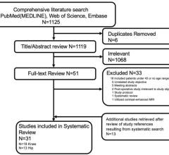
April 5, 2024 — Osteoarthritis — not age — may play the greatest role in determining the utility of MRI for patients 45 ...
April 05, 2024 Sponsored Content
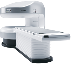
Fujifilm’s APERTO Lucent is a 0.4T mid-field, open MRI system addressing today’s capability and image quality needs ...
September 25, 2024 
News | Radiology Business
April 4, 2024 — Fujifilm Healthcare Americas Corporation, a leading provider of diagnostic and enterprise imaging ...
April 04, 2024 
News | Molecular Imaging
March 29, 2024 — Magnetic resonance imaging (MRI) is a cornerstone in the landscape of medical diagnostics, celebrated ...
March 29, 2024 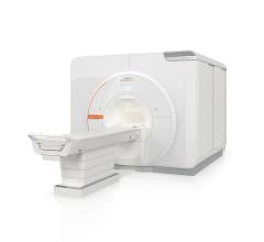
March 27, 2024 — Siemens Healthineers announces the Food and Drug Administration (FDA) clearance of the MAGNETOM Terra.X ...
March 27, 2024 Sponsored Content

Case Study | Radiology Imaging
In June, the Philips Radiology Experience Tour hit the road to provide healthcare professionals with an opportunity to ...
September 19, 2023 
News | FDA
March 27, 2024 — SyntheticMR announced that its next-generation imaging solution, SyMRI 3D, has received FDA 510(k) ...
March 27, 2024 
News | Artificial Intelligence
March 1, 2024 — Royal Philips, a global leader in health technology, and magnetic resonance imaging (MRI) software ...
March 01, 2024 
February 28, 2024 — Royal Philips, a global leader in health technology, today announced a significant milestone as it ...
February 28, 2024 Sponsored Content
Videos | Radiology Imaging
This summer, the Philips Radiology Experience Tour has been bringing Philips imaging modalities directly to the ...
August 14, 2023 
Hyperfine to Collaborate with Athletic Heart to Provide Brain Health Imaging for Former Pro Athletes
February 21, 2024 — Hyperfine, Inc., a groundbreaking health technology company that has redefined brain imaging with ...
February 21, 2024 
February 16, 2024 — Fujifilm Healthcare Europe will be presenting the Echelon Synergy – a revolutionary MRI scanner for ...
February 16, 2024 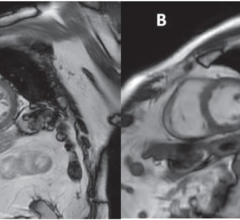
News | Cardiac Imaging
February 12, 2024 — According to the American Journal of Roentgenology (AJR), free-breathing cine-deep learning (DL) may ...
February 12, 2024 Sponsored Content

Case Study | Enterprise Imaging
The healthcare industry faces many different types of obstacles in today’s challenging marketplace. Staff shortages ...
August 01, 2023 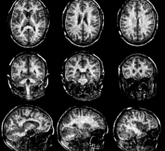
February 9, 2024 — Multiple sclerosis (MS) is a neurological disease that usually leads to permanent disabilities. It ...
February 09, 2024 
January 26, 2024 — InkSpace Imaging, a leader in innovative diagnostic medical device technology, is proud to announce ...
January 26, 2024 
News | Radiology Business
January 25, 2024 — Esaote Group, a leading Italian innovator in medical imaging, today unveiled its new brand identity ...
January 25, 2024 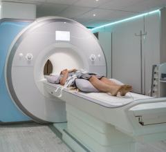
January 24, 2024 — Whole Body MRI, a private clinic committed to delivering accurate and comprehensive diagnostic ...
January 24, 2024 
News | Artificial Intelligence
January 23, 2024 — Quibim announced it has added an industry-leading cancer detection capability to its prostate tool ...
January 22, 2024 
January 18, 2024 — Abbott announced that the U.S. Food and Drug Administration (FDA) has approved expanded MRI labeling ...
January 18, 2024 
January 15, 2024 — MRI simulation software leader ScanLabMR announced the release of UltraLab, a breakthrough software ...
January 15, 2024 
January 11, 2024 — On January 9th, 2024, a scientific prototype for MRI-guided proton therapy was inaugurated in Dresden ...
January 11, 2024 © Copyright Wainscot Media. All Rights Reserved.
Subscribe Now