Sep. 15, 2025 —ScanLabMR has introduced its Signal-to-Noise Ratio (SNR) calculator. This feature gives MRI technologists ...
Magnetic Resonance Imaging (MRI)
MRI creates images from the magnetic resonance created in hydrogen atoms when they are polarized and an electromagnetic pulse is used to knock them off axis. This section includes MR analysis software, MRI scanners, gadolinium contrast agents and related magnetic resonance imaging accessories.

 September 18, 2025
Sept. 15, 2025 — Cook Medical and Siemens Healthineers have announced a strategic commercial partnership aimed at ...
September 15, 2025 
Sept. 10, 2025 —GE HealthCare announced it has entered into an agreement to acquire icometrix, a company focused on ...
September 10, 2025 Sponsored Content
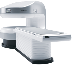
Fujifilm’s APERTO Lucent is a 0.4T mid-field, open MRI system addressing today’s capability and image quality needs ...
September 25, 2024 
News | Computed Tomography (CT)
Aug. 26, 2025— Esaote North America, Inc., a provider of dedicated MRI, Ultrasound, and Healthcare IT solutions, has ...
August 27, 2025 
Aug. 27, 2025 — Bayer has announced that a New Drug Application (NDA) for its investigational contrast agent ...
August 27, 2025 
News | RSNA 2025
Aug. 13, 2025 — Registration is now open for the RSNA 111th Scientific Assembly and Annual Meeting, the world’s leading ...
August 13, 2025 Sponsored Content

Case Study | Radiology Imaging
In June, the Philips Radiology Experience Tour hit the road to provide healthcare professionals with an opportunity to ...
September 19, 2023 
News | Radiology Imaging
Aug. 12, 2025 – Medical imaging methods such as ultrasound and MRI are often affected by background noise, which can ...
August 12, 2025 
July 31, 2025 —Hyperfine, Inc. has announced the enrollment of the first patients in the PRIME (Portable Rapid Imaging ...
July 31, 2025 
July 17, 2025 – Hyperfine, Inc.recently announced the first commercial sales of the next-generation Swoop system.The ...
July 24, 2025 Sponsored Content
Videos | Radiology Imaging
This summer, the Philips Radiology Experience Tour has been bringing Philips imaging modalities directly to the ...
August 14, 2023 
News | Artificial Intelligence
July 22, 2025 — GE HealthCare has topped a U.S. Food and Drug Administration (FDA) list of AI-enabled medical device ...
July 23, 2025 
July 16, 2025 — A scientific team supported in part by the National Institutes of Health (NIH) has developed a new ...
July 22, 2025 
News | Prostate Cancer
July 16, 2025 — Artificial intelligence can improve diagnostic consistency and reduce false-positives in prostate cancer ...
July 22, 2025 Sponsored Content

Case Study | Enterprise Imaging
The healthcare industry faces many different types of obstacles in today’s challenging marketplace. Staff shortages ...
August 01, 2023 
July 2, 2025 — Philips has received FDA 510(k) clearance for SmartSpeed Precise1 MR’s latest deep learning ...
July 03, 2025 
June 26, 2025 — Siemens Healthineers has received Food and Drug Administration clearance for the Magnetom Flow.Ace, its ...
June 26, 2025 
News | Prostate Cancer
June 26, 2025 – Quibim, a global provider of quantitative medical imaging solutions, has launched AI-QUAL, a new feature ...
June 26, 2025 
June 24, 2025 —Smart Soft Healthcare has announced that CoLumbo, the company's advanced AI spine assistant, has received ...
June 24, 2025 
May 29, 2025 — Hyperfine, Inc., producer of the world’s first FDA-cleared AI-powered portable MRI system for the brain — ...
May 29, 2025 
May 12, 2025 — GE HealthCare recently unveiled Signa Sprint, an FDA 510(k) pending[1] ultra-premium wide bore 1.5T high ...
May 12, 2025 
News | Pediatric Imaging
April 10, 2025 — Cincinnati Children’s and GE HealthCare will form a strategic research program focused on driving ...
April 10, 2025 
March 18 — Perspectum has released the results of a multinational study showing that LiverMultiScan can reduce liver ...
March 19, 2025 © Copyright Wainscot Media. All Rights Reserved.
Subscribe Now