
May 8, 2019 — Carestream introduced its ImageView Software Platform Windows 10 operating system to deliver enhanced ...
Advanced Visualization
Software used to manipulate or enhance CT and MRI datasets, including MPR, and 3-D (3D) image reconstruction, perfusion imaging, 3-D printing, and procedural planning and procedural navigation.

News | Medical 3-D Printing
May 3, 2019 — Bioengineers have cleared a major hurdle on the path to 3-D printing replacement organs with a ...
 May 03, 2019
News | Advanced Visualization
April 26, 2019 — Pickup Family Neurosciences Institute at Hoag in Newport Beach, Calif., announced the addition of the ...
April 26, 2019 Sponsored Content

Feature
In radiology departments, leveraging data and actionable knowledge are essential for making strategic clinical ...
November 16, 2020 
Videos | Cardiac Imaging
Dee Dee Wang, M.D., director of structural heart imaging, Henry Ford Hospital, Detroit, Mich., explains how patient ...
April 26, 2019 
Feature | Artificial Intelligence | By Greg Freiherr
Artificial intelligence (AI) may powerfully influence women’s health. Two vendors at the Society for Breast Imaging (SBI ...
April 22, 2019 
News | Advanced Visualization
April 4, 2019 — Increasing demand for innovative diagnostic techniques, neurological disorders and increasing disease ...
April 04, 2019 Sponsored Content

Blog | Advanced Visualization
Rural America does not show up in any atlas but, geographically, it accounts for 80 percent of the United States’ land ...
July 19, 2019 
Technology | Advanced Visualization
April 2, 2019 — Medical imaging and visualization company Medivis announced the launch of AnatomyX, its augmented ...
April 02, 2019 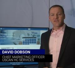
Sponsored Content | Videos | Advanced Visualization
GE Healthcare goes beyond core equipment maintenance to help clients solve some of their most important asset and ...
April 01, 2019 
News | Enterprise Imaging
March 29, 2019 — Medical imaging software company Novarad announced that it has appointed Paul Jensen as company ...
March 29, 2019 Sponsored Content
Videos | Advanced Visualization
GE Healthcare goes beyond core equipment maintenance to help clients solve some of their most important asset and ...
April 01, 2019 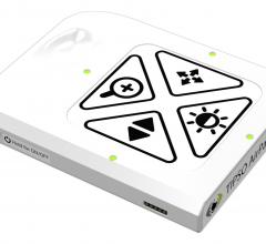
News | Interventional Radiology
March 25, 2019 — NZ Technologies Inc. announced the first published clinical review on its TIPSO technology’s ability to ...
March 25, 2019 
News | Advanced Visualization
March 18, 2019 – DrChrono Inc. and 3D4Medical have teamed up so practices across the United States can access 3-D ...
March 18, 2019 
Feature | Cardiac Imaging | By Greg Freiherr
Virtual reality (VR) and its less immersive kin, augmented reality (AR), are gaining traction in some medical ...
March 17, 2019 Sponsored Content
Videos | Information Technology
In this video Johann Fernando, Ph.D., Chief Operating Officer of FUJIFILM Medical Systems U.S.A., Inc. discusses his ...
February 07, 2019 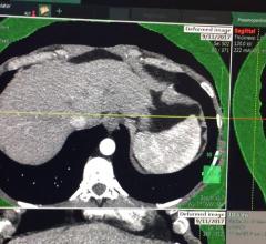
Videos | Advanced Visualization
This is an example of a new endoscopic virtual peritoneal inflation tool on the patient's computed tomography (CT) ...
March 05, 2019 
Videos | Orthopedic Imaging
This is an example of a 3-D printed pelvis that had multiple hip fractures and a second printed pelvis is from a post ...
March 05, 2019 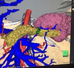
Videos | Advanced Visualization
This is an example of a new endoscopic 3-D imaging simulator created from a patient's computed tomography (CT) scan ...
March 05, 2019 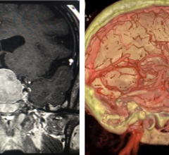
News | Radiology Imaging | By Melinda Taschetta-Millane
Here is the list of the most popular articles and videos on the Imaging Technology News (ITN) magazine website from the ...
March 04, 2019 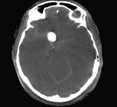
Technology | Angiography
March 1, 2019 — iSchemaView announced the release of RAPID Angio, a complete neuroimaging solution for the angiography ...
March 01, 2019 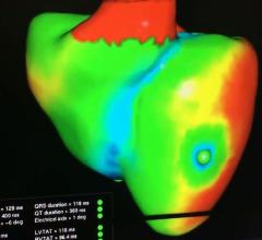
Videos | Cardiac Imaging
This is a virtual heart with the same electrophysiology characteristics as the real patient unveiled by Siemens at the ...
February 27, 2019 
Technology | Advanced Visualization
February 27, 2019 — Philips announced the launch of IntelliSpace Portal 11, the latest release of the company’s ...
February 27, 2019 
News | Advanced Visualization
February 25, 2019 — Philips will unveil a new mixed reality concept developed together with Microsoft that the company ...
February 25, 2019 © Copyright Wainscot Media. All Rights Reserved.
Subscribe Now