Electromagnetic therapies currently used in the National Aeronautics and Space Administration (NASA) space centers, including pulsed electro-magnetic field (PEMF), direct current, capacitive coupling, shock therapy and laser, to name a few, have been used widely for tissue regeneration in spinal fusion and fracture healing since the 1990s. Recent interest in application to inflammatory disorders is being internationally validated with the new generation of image guided treatments due to the long COVID pathologies that are ongoingly encountered. The possible relationship of autoimmune disease to oncologic processes is being explored by the immunologic and oncologic communities.
Microvascular Physiology
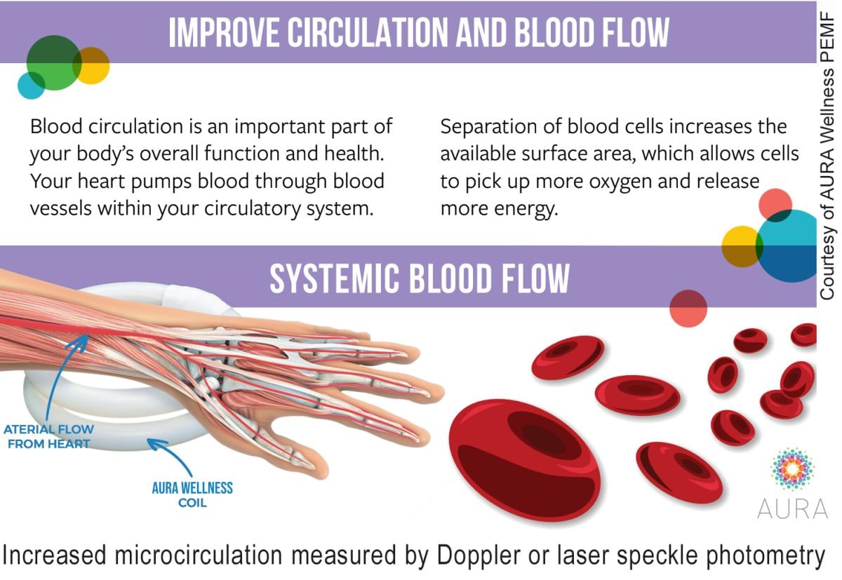
The smooth muscle content of vascular structures responds to the hormonal and autonomic nervous system control and is sensitive to electromagnetic fields (EMF). Blood flow is measurable in the microvessels (less than 150 microns) by Doppler ultrasound, speckle laser photometry, optical coherence tomography, intravital microscopy, dual photon-spectroscopy and reflective confocal microscopy, and may be studied noninvasively in real time, providing assessment of treatment effect and correlated with metabolic testing and functional genomics.
Blood distribution control mechanisms in the pre- and post-capillary microcirculation depends on the vessel caliber and have 3-5 vasomotions/minute, whereas larger vessels upstream and downstream have 1 vasomotion/minute. The faster vasomotion of the smallest vessels takes place autorhythmically while the larger vessels are controlled by the hormonal and/or autonomic nervous system. Aging effect and disease status reduces the frequency on vasomotion while certain signal configurations carried on the electromagnetic wave increase the vasomotion thereby increasing blood flow, tissue oxygenation, venous return and glymphatic clearance of waste products supporting tissue regeneration and immunologic activity. This noninvasive technology is widely used for diabetic disease of the eye and foot, and inflammatory skin disease, and was FDA approved for fracture healing over 30 years ago.
Inflammatory Dermatitis/Intestinal Inflammation
Generally considered autoimmune diseases, electromagnetic therapy increases venous flow out of the microvascular networks, extends the plasma distribution in the capillary network and increases spontaneous arteriolar vasomotion resulting in better regional oxygenation. Parallel changes of the subcutaneous tissue and intestine occur with functional electromagnetic treatment.1
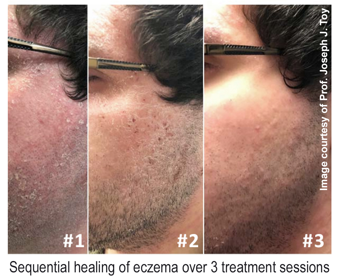
The use for psoriasis began about 40 years ago in Italy.2 The immune system response to tissue injury involves homeostasis and cell signaling that affects the rate of healing and scarring as stem cells are important in tissue regeneration.3 Low frequency EMF induces changes in cell proliferation, membrane structure and function, protein phosphorylation and adenosine triphosphate (ATP) synthesis.4 Successful regeneration requires an immune response that accurately polarizes immune cells so they balance between pro-inflammatory and pro-regenerative cell types. When inflammation becomes chronic multipotent mesenchymal stem cells (MSCs) modulate the recovery process.5 Healing involves cellular debris removal, activation of progenitor cells, immune modulation, angiogenesis and innervation of the regenerating tissue.6 Electromagnetic induction dosimetry incudes frequency, intensity and duration/time of exposure to balance the pro- and anti-inflammatory signaling processes of IL-1 beta secretion important in type 2 diabetes and other inflammatory states.7 EMF acts on calcium concentrations and Na/K pathways in humans8 and modulates endogenous electrical potential in plants, animals and humans.9 Continuous anecdotal reporting of the salutary effects of EMF are likely due to the cellular effect on homeostatic mechanisms10 and improvement in gut health seems due to decreased sticking of red blood cells and increased flow of white blood cells to penetrate into smaller capillaries.

Diabetic Macular Edema
Hyperglycemia and oxidative stress are the basis of diabetic retinopathy as the cell membrane does not transmit glucose/sorbitol which leads to tissue accumulation of advanced glycation end products with increased local oncotic pressure that damages connective tissue and produces vascular rigidity. The resulting hypoxia damages cell membranes, denatures protein, damages DNA and stimulates local angiogenesis by activating vascular endothelial growth factor (VEGF). Long-term hyperglycemia increases capillary permeability, blood viscosity and thrombocyte aggregation reducing retinal oxygenation. White blood cell leukocytes adhere more closely to the endothelium potentially producing capillary occlusion and damage the vascular endothelium leading to turbulent intravascular flow patterns starting in the middle periphery of the retina. As ischemia progresses the perifoveolar tissue is degraded. Intraocular pressure changes alter perfusion causing lowered pressure resulting in venous dilation further producing exudates, vessel wall weakening and microaneurysms.11 These aberrations correlate with optical and hemodynamic documentation.
Neuropathic Disorders
Various multidimensional signal devices (transcranial magnetic stimulation, repetitive transcranial electromagnetic stimulation, pulsed electromagnetic therapy) applied to the magnetic field are used in multiple sclerosis (MS)12 and Parkinson’s disease with alleviation of motor and non-motor symptoms.13 Studies have shown response of magnetic stimulation technologies combined with biofeedback or thermo-optic energy treatment to produce alteration in the retinal circulation and optic nerve.14
Doppler Applications
Blood vessel mapping using the various Doppler flow modalities is routinely used in both cancer treatment and reconstructive preoperative planning. In cancer surgery, it is important to identify aberrant large veins or significant arteries in the operative site so that hemostasis is maintained and postoperative blood loss is minimized. Before initiating cosmetic procedures or aesthetic treatments many plastic surgeons routinely perform a screening overview scan of the facial tissues including the eye, nose, jaw and neck to check for forgotten fillers or post procedure complications, such as subdermal scar formation, intradermal calcific deposits from healed previous injuries or retained silicone and other fillers that may have been injected in the past. Particular attention is focused on the nasal glabellar area because instances of total, permanent blindness have been occurring for over 10 years due to the inadvertent deposition of injectable filler or fat transplant material into the draining veins that supply the back of the eye. Advance warning of this danger zone means injectables may be deposited in a safer location. Fat transplants and silicone pads around the orbit occasionally put pressure on the venous return resulting in tissue discoloration and edema from venous obstruction while emboli to the ophthalmic arteries cutting off blood supply to facial regions cause sloughing of the affected ischemic skin. Vascular occlusion or insufficiency may be documented by Doppler studies and successful therapeutic relief documented in real time. Advanced 3-D Doppler systems allow for histogram vessel density measurement of neoplastic angiogenesis. (See Figures 1A, 1B and 1C.)
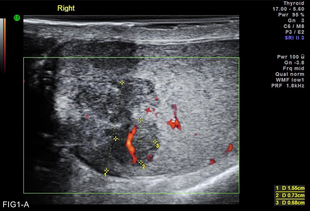
Fig 1-A: Power Doppler of 15 mm inflammatory testicular nodule
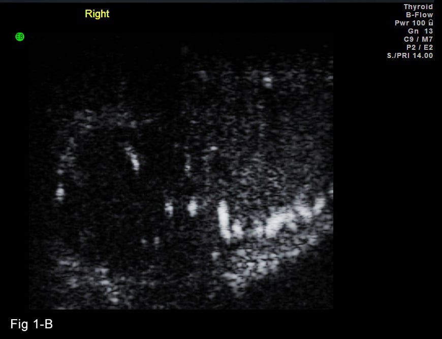
Fig 1-B: Microcirculation of inflammatory nodule with B flow ultrasound showing 100 micron vascularity
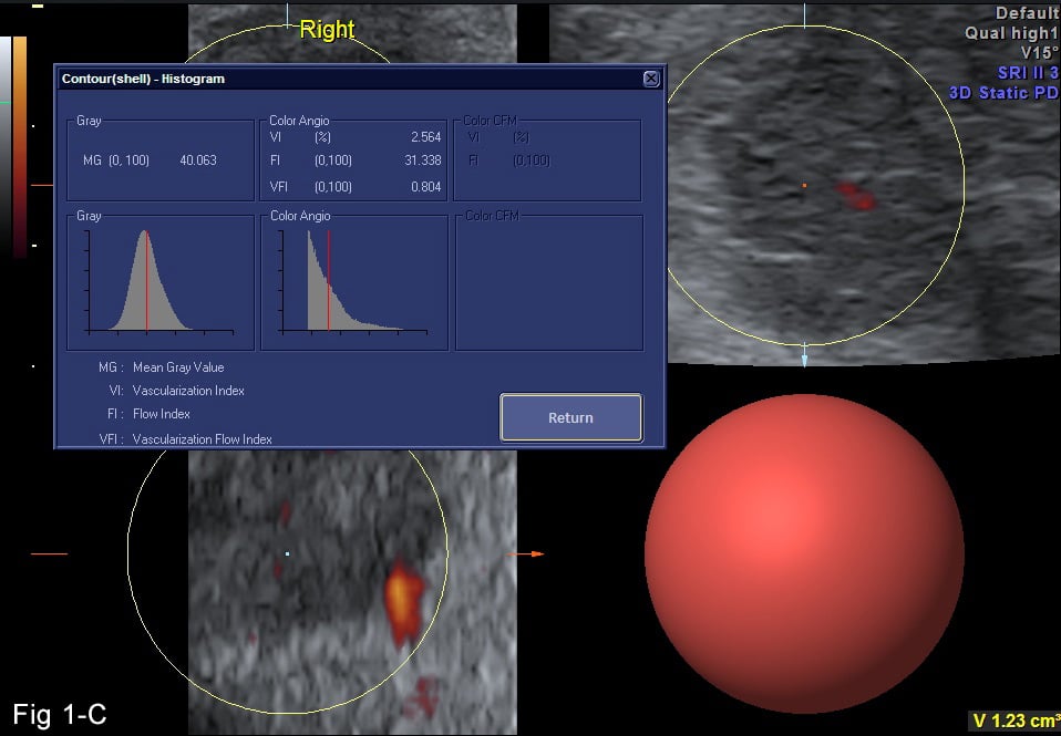
Fig 1-C: 3D Doppler histogram demonstrating inflammatory neovascular density of 2.5% of focal lesion controlled by electromagnetic field therapy for 6 months
This baseline neovascularity is used as a treatment surrogate endpoint allowing for image guided clinical reevaluation and timely correction of therapy. (See Figures 2A, 2B and 2C.)¹⁵
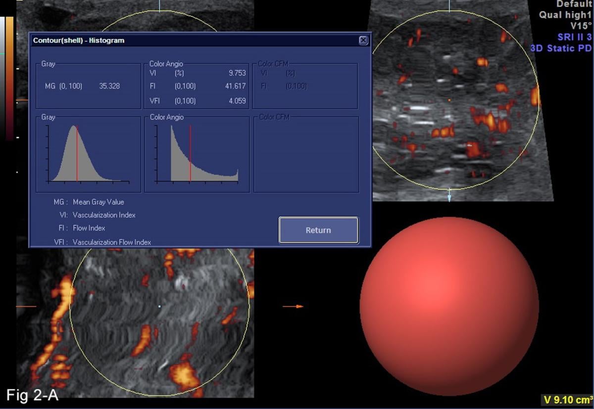
Fig 2-A: 3D Doppler histogram of testicular cancer pretreatment vessel density 9.7%
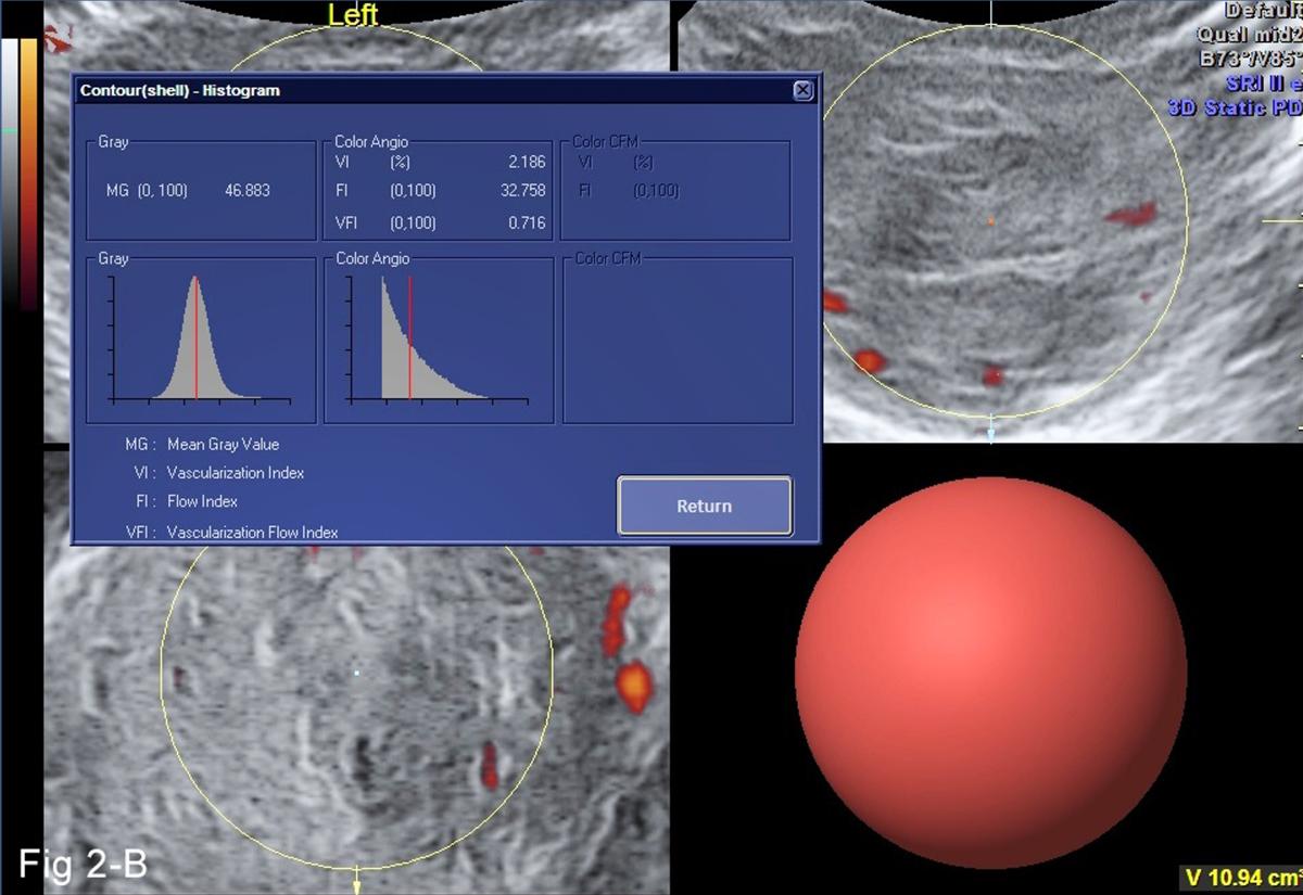
Fig 2-B: 4 months post EMF treatment documenting vessel density decrease from 9% to 2%
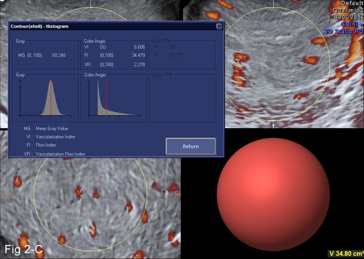
Fig 2-C: 6 month interval scan after treatment stopped demonstrating tumor neovascular increase
Orchietectomy followed this recurrence documentation
Contrast Enhanced Ultrasound
In 1990, Rodolfo Campani, MD, director of the University of Pavia Radiology Department in Italy, developed ultrasound contrast enhanced cancer imaging for liver tumors whose success was quickly followed by Drs. David Cosgrove, MD, (London) Nathalie Lassau, MD, (Paris) and Carlo Catalano, MD, (Naples) for malignancies of the breast, prostate and melanoma. Contrast enhanced ultrasound (CEUS) is currently used worldwide but not fully FDA approved in the United States. Microbubbles show tumor neovascularity with exquisite detail and are used to evaluate therapeutic response in solid organ disease. This contradicts standard RECIST studies demonstrating tumor enlargement during treatment may be related to apoptotic cell death with cystic degeneration or immune cell infiltration destroying malignant tissue. Co-existing benign disease may be indistinguishable from and integrated into the tumor mass. Doppler ultrasound or CEUS reliably verifies decreased angiogenesis in these cases instead of using contrast CT or DCE-MRI for confirmation. Thermal treatments such as cryotherapy, high-intensity focused ultrasound (HIFU), photodynamic treatment or laser ablation are designated completed when perfusing cancer arteries are no longer visible with the imaging criteria.
Intravital Microscopy and Advanced Optics
The small vessels supplying oxygen and removing tissue waste have specialized flow parameters observed in real time dedicated instrumentation validating smooth intravascular flow and rhythmic contractions of the endothelium. As high lipids and elevated blood viscosity with erythrocyte aggregation alter the vessel physiology white micro cholesterol debris appears. Normal vasomotion is 4-10 cycles per minute (cpm) is reduced in microneuropathy and problematic vascular flaps. This is prognostic data for cardiovascular disease but useful in evaluating burns, inflammatory dermatitis and collagen disorders, and will prove useful in diabetic neuropathy. Healing blood flows have high systolic and low diastolic values used for documenting fracture union for 25 years so a similar ratio could be applied on a microcirculatory level that is predictive of successful wound repair that balances cytokine effect to regenerate tissue while restricting fibrosis. Vascular physiology of aggressive cancers demonstrate abnormally low systolic resistive index vessels and a study on inflammatory dermatitis may be considered using a similar rationale.
Autoimmune Disease and Cancer
Abnormal immune responses that initially appear in the skin are associated with increased cancer incidence. Inflammatory vessels in psoriasis and infection are visibly cataloged since successful treatment is quantified by measured decrease in the number and type of abnormal vessels. High vascular immune vessel density is proportional to increased risk of future neoplastic tissue manifestations. Many arthritic conditions have coexistent dermal components alerting us to the probability of more extensive subclinical joint involvement. Botanical and cosmeceutical formulations have ameliorated many inflammatory diseases and possibly prevented malignant transformations and their therapeutic effect may be monitored before more aggressive approaches are initiated. International bio energy organizations are adopting the moniker “electroceuticals” to fit in with current academic trends.
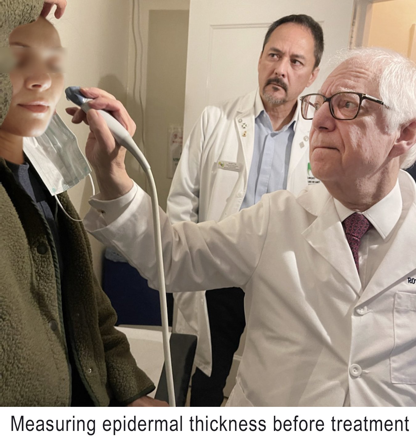
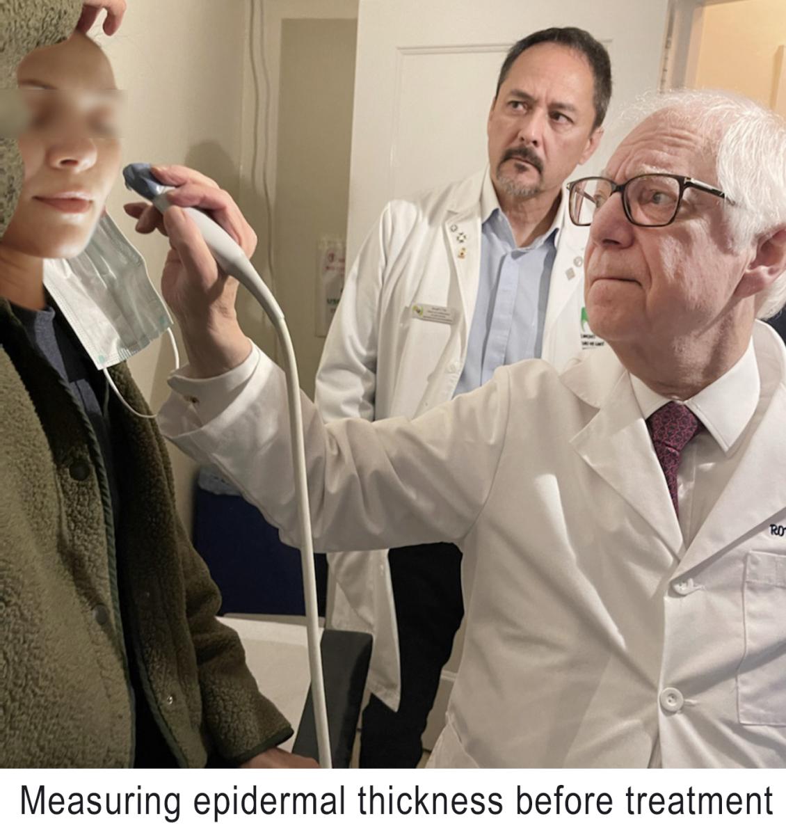
Subdermal imaging affords targeted access to inflammatory disease with the manifestation of subcutaneous vascular effects such as the “sun sign” of morphea¹⁶ and vasculitis of microarteries. Clinically lesions are variable and change with chronicity producing pain and skin tension effects. Histological diagnosis depends on the sample size, depth and biopsy site. Ultrasound equal or greater than 15 MHz reliably depicts increased epidermal and dermal thickness, decreased dermal echogenicity along with increased subdermal echogenicity and loss of the border between the dermis and subcutaneous tissue as useful monitor parameters for active disease. Spectral color Doppler shows increased flow dermal and subdermal flows as well as increased systolic arterial velocity possibly related to perivascular inflammation. The most reliable markers are increased subcutaneous echogenicity and increased cutaneous blood flow. While the “sun sign” of increased perivenous echogenicity has been associated with morphea activity, it is anecdotally present in other inflammatory diseases such as psoriasis, rosacea and rheumatoid arthritis. Optimal visualization of the echogenic halo includes normal subcutaneous echogenicity signifying that incidental perilesional observation of the “sun sign” indicates early aggressive systemic disease related to the dermal location suggesting a change in treatment options.
Dermal Elastography
Advances in the physical elasticity properties of tissue were first investigated by the Japanese in malignant melanoma and soft tissue sarcomas in 1990 since cancers are firm and benign lesions are soft and pliable. Strain elastogram verifiable results led to more accurate and quantifiable shear wave data replacing tissue biopsies in tumors of the Achilles tendon, breast, thyroid, liver, pancreas, testicle and prostate in many countries.
Robert L. Bard, MD, PC, DABR, FASLMS, is an active medical director of Cancer Diagnostic Imaging Center (NYC), using advanced 3-D ultrasonographic Doppler imaging to detect cancers in breast, prostate, skin, thyroid, melanoma and other areas. He has extensive credentials as a clinical researcher/validator and has published medical textbooks and science journals. He is also a member of the ITN Editorial Advisory Board.
References:
1. Mikrozirculation im Focus der Forschung ISBN 978-3-033-01464-0; 421-424, 2008
2. Castelpietra R etal [Initial experiences in the treatment of psoriasis with pulsating magnetic fields] Minerva Med 75: 2381-2387, 1984
3.
Ross CL etal The use of Pulsed Electromagnetic Field to Modulate Inflammation and Improve
Tissue Regeneration Bioelectricity 1:247-259, 201R9
4. Bassett C Fundamental aspects of therapeutic uses of pulsed electromagnetic fields [PEMF] Crit Rev Biomed Eng 17:451-529, 1989
5. Zhange J etal Cytokines, inflammation and pain Int Aneth Clin 45:27-37, 2007
6. DelaRosa O etal Toll-like receptors as modulators of mesenchymal stem cells Front Immunol 3;1-8, 2012
7. Eggenhofer E Mesenchymal stem cell-educated macrophages Transplant Res 1784:119-126, 2012
8. Pchelintseva E etal Mesenchymal control by calcium-activated potassium channels J Cell Physiol 233;3755-3768, 2017
9. Wade B A review of PEMF mechanisms at a cellular level Am J Health Res 1:51-55, 2013
10. Rosado M etal Immune modulating perspective for low frequency electromagnetic fields in innate immunity Front Public Health 6:85, 2018
11. Pilot study from Konsensus Konferenz Freudenstadt 2010
12. Ross CL etal The effect of low frequency EMF on mesenchymal stem cell differentiation Stem Cell Res 15:96-108, 2015
13. Vadala A etal, Mechanisms and therapeutic applications of electromagnetic therapy in Parkinson’s disease Behav Brain Funct 11;26-31, 2015
14. Bard R etal 3D Doppler imaging of the autonomic nervous system with EM Fields on retinal vessels Proceedings of the 2023 AIUM
15. Neuer A etal The role of heat shock proteins in reproduction Hum Reprod Update 6:149-159, 2000
16. Wortsman X Sunsign in morphea J Ultrasound Med 41:1817-1824, 2021
Related Ultrasound Imaging Content:
Ultrasound Systems Market Forecast is Strong for 2023 and Beyond
Ultrasound Systems Market Trends
2021 Review: Transcranial Doppler Ultrasound Imaging
Stroke Scans Could Reveal COVID-19 Infection
Lung Scans for Stroke Patients Could Provide Earlier COVID-19 Detection
PHOTO GALLERY: How COVID-19 Appears on Medical Imaging
Digital Health Devices Used at Point of Care May Improve Diagnostic Certainty
Insights on the Worldwide Handheld Ultrasound Imaging Devices Industry

 July 28, 2026


