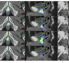
June 15, 2022 — Current guidelines used to plan salvage radiation treatments in patients with local recurrence of ...
Prostate Cancer
News and new technology innovations concerning how imaging technology can help diagnose and treat prostate cancer can be found on this channel.

News | Prostate Cancer
June 10, 2022 — GE Healthcare provides cutting-edge molecular imaging solutions that enable and increase access to ...
 June 10, 2022
 News | PET Imaging
June 8, 2022 — Blue Earth Diagnostics, a Bracco company and recognized leader in the development and commercialization ...
June 08, 2022 Sponsored Content
Videos | Prostate Cancer
Detecting metastatic disease early is key. Sand Lake Imaging, Florida, provides great value to both patients and ...
May 26, 2015 
News | Focused Ultrasound Therapy
May 12, 2022 — Henry Ford Health is the first in Michigan to offer Robotic High Intensity Focused Ultrasound (HIFU) for ...
May 12, 2022 News | Digital Pathology
April 27, 2022 — Lumea, a global leader in integrated digital pathology solutions and Verily, an Alphabet precision ...
April 27, 2022 
News | Prostate Cancer
April 25, 2022 — Over the past 15 years, public health authorities have downgraded recommendations for the prostate ...
April 25, 2022 
News | Prostate Cancer
April 19, 2022 — Blue Earth Therapeutics, a Bracco company and emerging leader in the development of innovative next ...
April 19, 2022 
News | Prostate Cancer
April 15, 2022 — Prostate cancer is the most common malignant tumor in men in Germany, with about 62,000 new cases ...
April 14, 2022 
News | PET-CT
April 11, 2022 — A novel nuclear medicine combination therapy has been proven safe and effective in men with heavily pre ...
April 11, 2022 
News | Prostate Cancer
April 11, 2022 — RSIP Vision, an experienced leader in driving innovation for medical imaging through advanced ...
April 11, 2022 
News | Prostate Cancer
Mount Sinai Health System Launches First-Ever Mobile Prostate Cancer Screening Unit in New York City
April 6, 2022 — On Friday, April 1 at 2 p.m., the Milton and Caroll Petrie Department of Urology at Mount Sinai will ...
April 06, 2022 
News | Coronavirus (COVID-19)
April 6, 2022 — Research in the March 2022 issue of JNCCN—Journal of the National Comprehensive Cancer Network examined ...
April 06, 2022 
News | Radiology Education
April 5, 2022 — The American College of Radiology (ACR) has selected 22 teams as the first cohort of the ACR Learning ...
April 05, 2022 
News | Prostate Cancer
March 28, 2022 — ITM Isotope Technologies Munich SE (ITM), a leading radiopharmaceutical biotech company, supports ...
March 28, 2022 
News | Prostate Cancer
March 23, 2022 — With today’s U.S. Food and Drug Administration (FDA) approval of 177Lu-PSMA-617—a radiopharmaceutical ...
March 23, 2022 
News | Prostate Cancer
March 23, 2022 — A new study from Keck Medicine of USC finds that the incidence rate of metastatic prostate cancer has ...
March 23, 2022 
News | Prostate Cancer
March 11, 2022 — NorthStar Medical Radioisotopes, LLC, a global innovator in the development, production and ...
March 11, 2022 
News | Prostate Cancer
February 18, 2022 — Blue Earth Diagnostics, a Bracco company and recognized leader in the development and ...
February 18, 2022 
News | Prostate Cancer
February 17, 2022 — An interim analysis of an ongoing Phase III study from UCLA Jonsson Comprehensive Cancer Center ...
February 17, 2022 
News | Radiology Imaging
The American College of Radiology (ACR) reports that several states are considering breast, colon and lung cancer ...
February 14, 2022 © Copyright Wainscot Media. All Rights Reserved.
Subscribe Now