
News | Computed Tomography (CT)
The Center for Diagnostic Imaging (CDI) in Miami recently released a list of important preparations patients should make before undergoing a computed tomography (CT) scan.
 September 05, 2018
News | PET Imaging
Eli Lilly and Co. and Avid Radiopharmaceuticals Inc. announced a Phase 3 study of positron emission tomography (PET) imaging agent flortaucipir F-18 met its two primary endpoints, defined as predicting brain tau pathology and predicting Alzheimer's disease diagnosis.
September 05, 2018 
News | Colonoscopy Systems
Check-Cap Ltd. announced the interim results for its post-CE approval study of the C-Scan system Version 3, an ingestible, capsule-based device for preparation-free colorectal cancer (CRC) screening. The company said data from the multicenter clinical investigation showed promising results for detecting patients with polyps in an un-prepped colon.
September 04, 2018 Sponsored Content
Sponsored Content
Videos | Radiology Business
Radiology departments have many different needs and face a wide variety of challenges that can impact their departments ...
November 11, 2025 
News | Vendor Neutral Archive (VNA)
Novarad Healthcare Enterprise Imaging has taken the highest rated spot on Gartner’s Peer Insights technology review platform, outranking every other competitor in the vendor-neutral archive (VNA) market category.
September 04, 2018 
News | Enterprise Imaging
September 4, 2018 — LifeImage announced that its recently launched application, LITE, has helped to dramatically ...
September 04, 2018 
News | Enterprise Imaging
Agfa HealthCare and Greenville Health System (GHS), South Carolina, announced the successful implementation of a comprehensive enterprise imaging system. The implementation will facilitate greater efficiencies in clinical operations, improve clinical confidence, and enable cost reduction through convergence of systems and timely access to medical images and diagnostic tools.
August 31, 2018 Sponsored Content
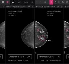
Feature | Breast Imaging
Despite decades of progress in breast imaging, one challenge continues to test even the most skilled radiologists ...
October 24, 2025 
News | Remote Viewing Systems
August 30, 2018 — New South Wales, Australia’s Newborn and pediatric Emergency Transport Service (NETS) recently ...
August 31, 2018 
News | Radiology Business
A new report from data and analytics company GlobalData suggests that Amazon is poised to make huge strides in healthcare as the Internet retail giant prepares to enter a new market. The report highlights a business model that facilitates easier connectivity between providers and patients, a powerful online platform and its artificial intelligence (AI) interface, Alexa.
August 31, 2018 
News | Neuro Imaging
A new, highly accurate magnetic resonance imaging (MRI) technique can monitor iron levels in the brains of multiple sclerosis (MS) patients. The technique could also help identify those at a higher risk for developing physical disability, according to a study published in the journal Radiology.
August 31, 2018 Sponsored Content
Sponsored Content
Videos | Radiology Business
Bayer Radiology’s Barbara Ruhland and Thom Kinst discuss how radiology departments can address the many different ...
October 09, 2025 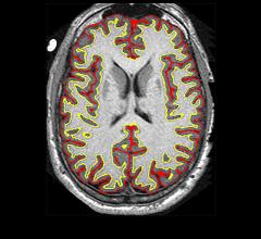
News | Neuro Imaging
August 30, 2018 — Results from a clinical trial of more than 250 participants with progressive multiple sclerosis (MS) ...
August 30, 2018 
News | Radiation Therapy
August 29, 2018 — Although the success or failure of radiation therapy for cancer has long been associated with the ...
August 29, 2018 
A newly developed rapid imaging protocol quickly and cheaply diagnosed heart ailments in patients in Peru, according to new research in Journal of the American Heart Association.
August 29, 2018 Sponsored Content
Case Study | PACS
eHealth Saskatchewan plays a vital role in providing IT services to patients, health care providers, and partners such ...
February 03, 2025 
Videos | Treatment Planning
A discussion with Kevin Moore, Ph.D., DABR, deputy director of medical physics and associate professor, University of ...
August 28, 2018 
News | X-Ray
August 28, 2018 — A new study has demonstrated a method that produces novel light beams from synchrotron light sources ...
August 28, 2018 
News | Neuro Imaging
iSchemaView announced that more than 575 stroke centers in 22 countries have selected the RAPID advanced imaging platform for cerebrovascular imaging, with 520 currently installed. RAPID technology assists physicians in the analysis of brain images using automated tools for CT ASPECTS, computed tomography (CT) angiography, CT perfusion, magnetic resonance (MR) diffusion and perfusion for more than 85,000 stroke cases per year. RAPID is also currently deployed in six multi-center clinical trials globally.
August 27, 2018 
News | SPECT Imaging
In the largest known brain imaging study, scientists from five institutions evaluated 62,454 brain single photon emission computed tomography (SPECT) scans to investigate factors that accelerate brain aging. The study — conducted by scientists from Amen Clinics (Costa Mesa, Calif.), Google, Johns Hopkins University, University of California Los Angeles and the University of California San Francisco — encompassed more than 30,000 individuals from 9 months old to 105 years of age.
August 27, 2018 
News | Artificial Intelligence
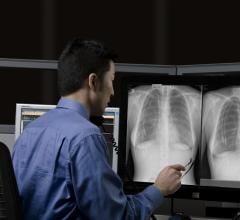
The Radiological Society of North America (RSNA) has launched its second annual machine learning challenge. The RSNA Pneumonia Detection challenge invites teams to develop algorithms to identify and localize pneumonia in chest X-rays.
August 27, 2018 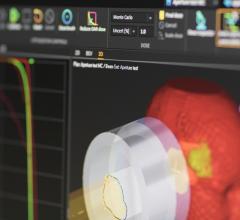
News | Treatment Planning
Tsuyama Chuo Hospital in Okayama Prefecture, southwest Japan, has commenced clinical use of RayStation to plan pencil beam scanning (PBS) treatments at its Proton Beam Cancer Center. On May 15, the center carried out the world’s first patient treatment using RayStation together with Mitsubishi Electric’s proton therapy system, with a plan created in RayStation 6.
August 27, 2018 
News | PET Imaging
August 24, 2018 — In a small study of military personnel who had suffered head trauma and had reported memory and mood ...
August 24, 2018 
News | Radiation Therapy
Children with certain types of brain tumors who undergo radiation treatment are less likely to recall the specifics of events occurring after radiation than to remember pre-treatment happenings, according to a Baylor University study. The study compared these children to children with healthy brains.
August 24, 2018 © Copyright Wainscot Media. All Rights Reserved.
Subscribe Now