Twenty-seven radiologists at Washington Radiology Associates (WRA), a large private imaging practice in the Washington, D.C., metropolitan area, perform 85,000 mammograms annually at six clinical offices. In 2011, WRA replaced all of its 2D mammography systems with 15 Hologic Selenia Dimensions systems.

Located in north central West Virginia – an area hit particularly hard by the economic recession – Davis Health System is comprised of a 90-bed community hospital, critical access hospital, community outreach clinics and related facilities. This forward-thinking organization has made big strides in putting its patients first by investing in medical imaging information technology from McKesson.

Soroka University Medical Center in Beer-Sheva, Israel, has successful experience using the Philips Myocardial Defect Assessment solution.


Positron emission mammography (PEM) is an imaging technique designed to provide metabolic visualization of lesions in the breast tissue. The Naviscan high-resolution PEM system is a dual-detector system consisting of two flat, high-resolution detector heads using lutetium yttrium oxyorthosilicate in a 2 x 2 x 13 mm size. The detectors are mounted inside mammogram compression paddles with a 17 x 24 cm field of view.  The paddles can be rotated to optimize imaging to match images acquired in standard mammograms.  This allows for direct comparison with mammogram images.


By focusing on the effective use of electronic health records (EHRs) with certain capabilities, the legislation regarding meaningful use (MU) makes it clear that the adoption of EHRs is not a goal in itself: It is the use of this technology to achieve health and efficiency goals that matters most. While many radiologists have opposed the one-size-fits-all approach to this incentive program, MU clearly impacts the radiology community and is here to stay.


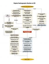
To achieve sustainable excellence in the quality and safety of healthcare, the radiology department at Broward Health Medical Center (BHMC) took a proactive approach to identify bottlenecks in study flows and streamline processes at every stage of imaging studies, from order entry to report posting. Its ultimate goal was to provide ordering physicians with radiologists’ reports in time to support a clinical diagnosis in an acute setting.


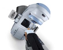
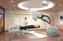
The external beam radiotherapy (EBRT) market has been active in recent months, with advances in techniques, devices and treatment planning software that help radiation oncology clinicians provide patients with the most targeted radiation delivery possible. With an estimated 1.1 million patients in the United States treated with radiation in 2009 (including both external beam and brachytherapy delivery), up 15 percent from 2007,[1] and worldwide growth in the radiation therapy market expected to grow at a compound annual rate of 9.1 percent by 2018,[2] activity in this sector can be expected to continue.


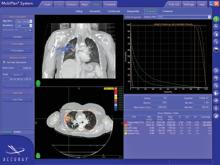
The preferred treatment for patients with operable lung cancer is surgical resection. However, many lung cancer patients whose tumors are technically resectable have other medical problems, such as chronic obstructive pulmonary disease, which make it difficult to endure lung cancer surgery. Until the last decade these patients were treated with conventional radiotherapy, but more recently stereotactic body radiotherapy (SBRT), or the accurate delivery of high doses of radiation over a short period of time, has emerged as an alternative treatment.


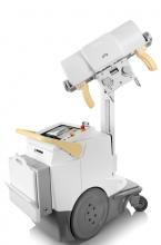
When it comes to modalities, why select digital radiography (DR)? There are several reasons. First, DR is fast. The speed inherent in the technology improves overall workflow for the entire radiology department. Also, image quality is superb and the required X-ray doses can be significantly lower than with traditional modalities.


Subscribe Now