December 7, 2021 — Xoran Technologies was recently notified of a grant award from the National Heart, Lung, and Blood Institute (NHLBI) of the National Institutes of Health (NIH) to support the company’s research and development efforts for lung computed tomography (CT) imaging.
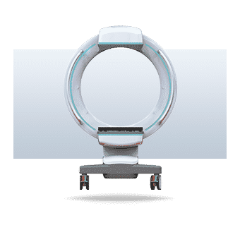
Leveraging its knowledge and experience in the field as the pioneer and medical market leader in cone beam CT, Xoran has proposed TRON—an open-bore, truly mobile CT to assist in the identification of lung disease. In addition, Xoran has established collaborative partnerships with specialists in Pulmonary and Critical Care Medicine and Radiology at the University of Michigan, Ann Arbor.
“We are grateful to NHLBI for giving us an opportunity to bring this new application to life,” stated William van Kampen, Xoran’s Chief Technology Officer and principal investigator on the research project. “We are excited to be working with the U-M team on a device that will allow point-of-care imaging of the lungs for helping patients who otherwise might have no good imaging option.”
With the xCAT IQ—an FDA 510k-cleared mobile CT system for bone and brain imaging, Xoran has shown capability in point-of-care (POC) CT solutions for the intensive care unit (ICU) and the operating room. This new TRON project allows Xoran to optimize a POC specifically for thoracic imaging for a variety of needs. The combined Xoran and U-M teams aim to develop a highly deployable CT scanner intended for use in the ICU, especially for patients with acute respiratory failure requiring mechanical ventilation, among other uses.
“It’s hard to overstate how transformative this technology would be for us in the ICU,” said Robert Dickson, MD, Associate Professor in Pulmonary & Critical Care Medicine, Deputy Director of the Michigan Center for Integrative Research in Critical Care, and a clinical collaborator at the University of Michigan. “Every day, we make clinical decisions based on chest X-rays, which are limited in what they can tell us about what is going on in the chest or abdomen. Our patients are often too sick to transport down to radiology, or they have a communicable disease like COVID-19 that we don’t want to spread around the hospital. A bedside scanner would have an immediate impact on how we manage our sickest patients.”
That’s not everything that Xoran aims to achieve with this grant project and the future TRON device.
“The TRON could help with rapid deployment for diagnostics in similar respiratory pandemic situations in the future. Really, it’s not just about the mobility; TRON will be easy-to use-and accessible,” explained Misha Rakic, Xoran’s Chief Executive Officer. “The goal is to offer a point-of-care CT device that is widely available and affordable to support diagnosis, triaging, and monitoring of respiratory and other patients who are too risky to move to centralized radiology. This paradigm could easily be applied to other modalities, and we can potentially provide a ‘one-stop shop’ POC solution for any CT and X-ray imaging need in the future.”
Xoran has a successful track record of receiving awards for commercializing SBIR technologies, such as the MiniCATTM CT scanner and a future device for spine CT imaging with integrated surgical navigation. Today, Xoran is an innovator and medical market leader with over 1,000 installations globally.
For more information: www.xorantech.com


 February 09, 2026


