December 15, 2014 — GE Healthcare’s Signa Pioneer, a new 510(k) pending 3.0T magnetic resonance imaging (MRI) system, offers a unique feature where it can create six imaging protocols from a single scan. The vendor said this will greatly reduce the amount of time required to image patients and enable an additional patient per hour to be scanned.
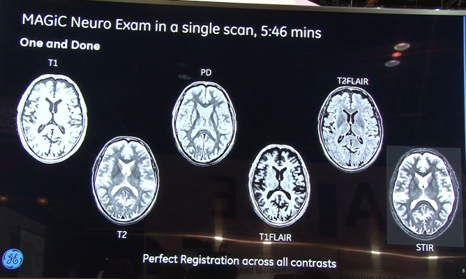
The system’s MAGiC software offers the ability to generate multiple image contrasts in a single MRI scan including T1, T2, STIR, T1 FLAIR, T2 FLAIR and PD weighted images of the brain in a single acquisition. Another innovation is that the contrast of images can be changed even after completing the scan by simply moving the cursor on the MAGiC interface to change acquisition parameters such as TR, TE and TI.
MAGiC, the result of a collaboration with SyntheticMR AB. With MAGiC, a single scan that delivers six contrasts can be completed in as little as one-third the total time taken to acquire each contrast separately using conventional techniques. This time saved could potentially allow clinicians to scan one more patient per hour.
The Signa Pioneer also uses GE’s enhanced SilentScan package, enabling nearly noiseless scans for musculoskeletal (MSK) imaging and spine imaging, in addition to a complete neuro exam that also includes diffusion weighted imaging (DWI). Conventional MRI scanners can generate noise in excess of 110 decibels (dBA) levels, roughly equivalent to a rock concert. SilentScan dramatically reduces the scan to just three dBA above ambient noise for most head exams, a major differentiator for patient comfort.
The Signa Pioneer also features technology called total digital imaging (TDI) enabling improved image quality and increasing signal to noise ratio (SNR) by up to 25 percent. TDI is composed of direct digital interface (DDI), digital surround technology (DST) and digital micro switching (DMS).
• DDI employs an independent analog-to-digital converter to digitize inputs from each of the 97 RF channels. Every input is captured and every signal is digitized.
• DST combines the digital signal from every surface coil element with the signal from the integrated RF body coil resulting in richer, higher SNR spine and body images with better homogeneity and uniformity.
• DMS is an advancement in RF coil design with intelligent micro electro-mechanical switches (MEMS) that support ultra-fast coil switching, which enables future expansion of Zero TE imaging capabilities. Zero TE enables imaging of tissues that are conventionally difficult to see with MRI, such as cortical bone, ligaments and tendons.

 June 12, 2026


