News | Prostate Cancer | July 22, 2025

An accepted manuscript published in the American Roentgen Ray Society’s American Journal of Roentgenology  looks at how artificial intelligence can improve diagnostic consistency and reduce false-positives in prostate cancer detection.

Lesion Identification by AI and Histopathologic Annotation

July 16, 2025 — Artificial intelligence can improve diagnostic consistency and reduce false-positives in prostate cancer detection, according to an accepted manuscript published in the American Roentgen Ray Society’s American Journal of Roentgenology (AJR).

Noting enhanced clinical utility for radiologists, “AI may enhance consistency and reduce false-positives in prostate biparametric MRI (bpMRI) interpretation,” clarified AJR corresponding author Baris Turkbey, MD, from the National Cancer Institute and National Institutes of Health in Bethesda, MD. “Further optimization is required to improve sensitivity without compromising specificity.”

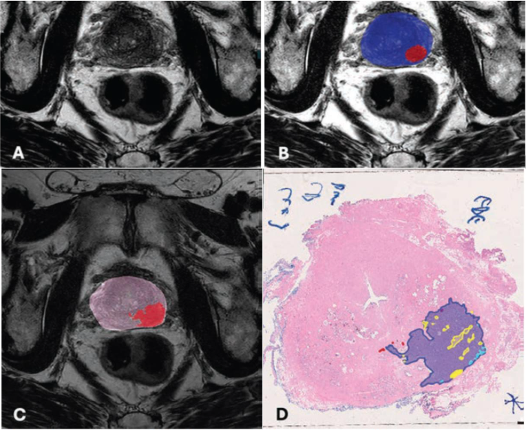
Turkbey et al.’s multireader, multicenter study used a balanced incomplete block design for MRI randomization. Six radiologists of varying experience interpreted bpMRI scans with and without AI assistance in alternating sessions. The reference standard for lesion-level detection for cases was whole-mount pathology after radical prostatectomy; for control patients, negative 12-core systematic biopsies. In all, 180 patients (120 case group, 60 control group) who underwent mpMRI and prostate biopsy or radical prostatectomy between January 2013 and December 2022 were included. The AJR authors then assessed lesion-level sensitivity, PPV, patient-level AUC for clinically significant prostate cancer and prostate cancer detection, and interreader agreement in lesion-level PI-RADS scores and size measurements.

Ultimately, AI-assisted bpMRI interpretation improved lesion-level PPV for prostate cancer (PI-RADS ≥ 3: 80.9% vs. 69.4%, p < .001) and significantly enhanced interreader agreement for PI-RADS scoring (κ = 0.748 vs. 0.336, p < .001) but slightly reduced sensitivity.

A supplement to this AJR manuscript is available online.


Lesion Identification by AI and Histopathologic Annotation
77-year-old man with serum PSA of 17.8 ng/mL. A) T2-weighted (T2W) MRI shows hypointensity in the left posterolateral peripheral zone. B) AI segmentation map overlaid on T2W image shows predicted prostate segmentation boundary in blue and PCa-suspicious lesion prediction segmentation in red. C) Ground truth histopathology slide registered to MR image shows annotated lesion segmented in red. D) Histopathologic annotation of whole mount prostate tissue after radical prostatectomy. This patient was found to have Gleason 4+3 disease in the region marked in blue. Yellow outlines represent cribriform architecture, red represents regions of Gleason 3, and light blue represents areas of lymphovascular invasion.


Related Content

News | Innovative Hospitals

May 27, 2026 — Nearly two years after announcing plans for a “real-world” academic-industrial collaboration, GE ...

Time June 03, 2026
arrow
News | Cardiac Imaging

May 21, 2026 — A team of researchers from Carnegie Mellon University, in collaboration with Cleveland Clinic’s ...

Time May 22, 2026
arrow
News

May 21, 2026 – Artera, the developer of multimodal artificial intelligence (MMAI)-based prognostic and predictive cancer ...

Time May 22, 2026
arrow
News | Radiology Imaging

May 18, 2026 — DICO, a company specializing in the creation of distributed diagnostic infrastructure for radiology, has ...

Time May 19, 2026
arrow
News | Imaging Software Development

April 28, 2026 — Avatar Medical has been granted FDA 510(k) clearance for Avatar Medical Vision, its software platform ...

Time April 28, 2026
arrow
News | Contrast Agents

April 23, 2026 — On April 23, GE HealthCare announced the first patient has been dosed in the international, multi ...

Time April 23, 2026
arrow
News | Radiology Imaging

April 20, 2026 — Bracco Imaging has announced a strategic alliance with NYU Langone Health to advance innovation in ...

Time April 23, 2026
arrow
News | Radiology Imaging

April 7, 2026 — Onvida Health and Siemens Healthineers have entered a 10-year Value Partnership¹ designed to bring the ...

Time April 09, 2026
arrow
News | Radiology Business

March 31, 2026 — Radon Medical Imaging, a medical imaging equipment maintenance and repair services company, has has ...

Time March 31, 2026
arrow
News | Radiology Imaging

March 26, 2026 — GE HealthCare has announced a renewed research collaboration with Stanford Medicine Department of ...

Time March 30, 2026
arrow
Subscribe Now