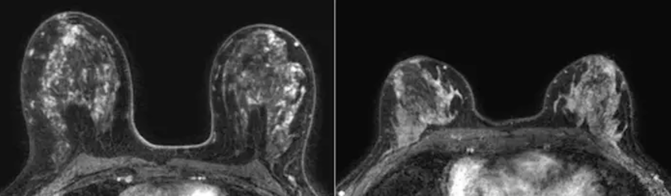
Representative transverse dynamic contrast-enhanced MRI scans in patients in the low and high tertiles of volume of enhancing parenchyma. Please note differences in high and low volumes of enhancing parenchyma are not visible to the naked eye on MRI scans. Left: Baseline MRI scan in a 50-year-old woman with a body mass index (BMI) of 23 and marked background parenchymal enhancement (BPE) who was stratified into the high tertile of volume of enhancing parenchyma. Cancer was detected with MRI in the second screening round. Right: Baseline MRI scan in a 51-year-old woman with a BMI of 24 and marked BPE who was stratified into the low tertile of volume of enhancing parenchyma. No cancer was detected during 6 years of follow-up. Image courtesy of RSNA.Br
August 9, 2023 — A machine learning model found that background parenchymal enhancement (BPE) on breast MRI is an indicator of breast cancer risk in women with extremely dense breasts, according to a study published in Radiology, a journal of the Radiological Society of North America (RSNA).
Women with extremely dense breasts are at a three- to six-times higher risk of developing breast cancer compared to women who have fatty breasts. Since mammography is less sensitive in detecting early-stage breast cancer in women with dense breasts, women between the ages of 50 and 75 years with dense breasts may benefit from additional MRI screening.
Another breast cancer risk factor is BPE, which is the level of normal fibroglandular tissue that enhances on breast MRI. However, not much is known about how BPE compares to other more established clinical risk factors of breast cancer, such as age, body mass index (BMI) family history, and breast density.
“Thus far, studies on breast cancer risk factors have typically focused on women at high lifetime risk of developing breast cancer,” said study co-author Kenneth G. A. Gilhuijs, Ph.D., from the Department of Radiology at the University Medical Center Utrecht in the Netherlands. “This is the first study that we know of that demonstrates an association between background parenchymal enhancement and occurrence of breast cancer in women with extremely dense breasts.”
To determine how much BPE is an indicator of breast cancer risk, the researchers used dynamic contrast-enhanced MRI exams from 4,553 participants in the Dense Tissue and Early Breast Neoplasm Screening (DENSE) Trial, a large multi-institutional study based in the Netherlands, to develop a deep learning model to automatically identify the fibroglandular tissue. The MRI exams were performed every two years in eight hospitals in the Netherlands between December 2011 and January 2016.
After adjusting for age, BMI and BPE, the researchers found that breast cancer occurrence was greater in women with higher volumes of enhancing parenchyma compared to women with low volumes of enhancing parenchyma.
Of the 4,553 women included in the study, 122 were diagnosed with breast cancer. Roughly 63% of them were diagnosed after the first round of screening. An average cancer detection time of 24 months was associated with the remaining women diagnosed with breast cancer.
“Parenchyma does not enhance uniformly on MRI,” Dr. Gilhuijs said. “This method calculates all the different subvolumes at which the parenchyma enhances and sorts them from high to low.”
The researchers point out that while the implementation of supplemental MRI screening in women with dense breasts will result in fewer interval cancers—which are breast cancers that are diagnosed in between routine mammography screenings—it will also further strain radiologist workloads. Developing more personalized strategies to deal with the added number of screenings may help alleviate the strain on the healthcare field.
“Our study is a first step in a direction to further tailor the frequency of supplemental MRI screening to individual women with dense breasts, focusing not only on breast density as a main risk factor but also on other properties of the breast established from a first screening MRI,” Dr. Gilhuijs said.
For more information: www.rsna.org
Find more RSNA23 conference coverage here
Related Breast Density Content:
VIDEO: FDA Update on the US National Density Reporting Standard - A Discussion on the Final Rule
One on One … with Wendie Berg, MD, PhD, FACR, FSBI
Task Force Issues New Draft Recommendation Statement on Screening for Breast Cancer
Creating Patient Equity: A Breast Density Legislative Update
AI Provides Accurate Breast Density Classification
VIDEO: The Impact of Breast Density Technology and Legislation
VIDEO: Personalized Breast Screening and Breast Density
VIDEO: Breast Cancer Awareness - Highlights of the NCoBC 2016 Conference
Fake News: Having Dense Breast Tissue is No Big Deal
The Manic World of Social Media and Breast Cancer: Gratitude and Grief
Related Breast Imaging Content:
Single vs. Multiple Architectural Distortion on Digital Breast Tomosynthesis
Today's Mammography Advancements
Digital Breast Tomosynthesis Spot Compression Clarifies Ambiguous Findings
AI DBT Impact on Mammography Post-breast Therapy
ImageCare Centers Unveils PINK Better Mammo Service Featuring Profound AI
Radiologist Fatigue, Experience Affect Breast Imaging Call Backs
Fewer Breast Cancer Cases Between Screening Rounds with 3-D Mammography
Study Finds Racial Disparities in Access to New Mammography Technology

 June 02, 2026


 Clearance")