Diffusion tensor imaging increases ability to remove benign tumors in children.
December 4, 2009 - Operative plans for removing juvenile pilocytic astrocytoma, or JPA, tumors in the thalamus of the brain can be augmented with diffusion tensor imaging, or DTI, according to a new study published in last week's issue of the Journal of Neurosurgery: Pediatrics.
Operating on patients with deep-seated tumors such as JPA, a benign tumor most frequently observed in children and young adults in the thalamus, remains a neurosurgical challenge. Conventional imaging techniques, such as structural MRI, has helped to reveal major anatomical features of the brain, primary gray matter.
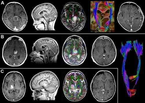
DTI, a variation of MRI, can help identify white matter, or nerve fiber bundles, using specific radio-frequency and magnetic field pulses to track the movement of water molecules of the brain. In most brain tissue, water molecules diffuse in all different directions. But they tend to diffuse along the length of axons, whose coating of white, fatty myelin holds them in. Scientists can create pictures of axons by analyzing the direction of water diffusion.
The sensitivity of DTI imaging allows for the visualization of nerve fiber bundles in the brain. This information can maximize the potential of completely removing the tumor, while avoiding damage to the fiber bundles that are directly related to motor functions of the patient.
“This study of six children with thalamic JPA showed that using advanced MRI technology can help identify distorted nerve fiber bundles around brain tumors,” said Jeffrey H. Wisoff, M.D., director of the Division of Pediatric Neurosurgery at NYU Langone Medical Center. “This allows an otherwise inoperable tumor to be completely removed which can hopefully lead to a cure.”
The co-authors of the study include Yaron Moshel, M.D., Ph.D., of the department of neurosurgery, David J. Monoky, M.D., clinical assistant professor in the department of radiology and Robert E. Elliott, M.D., in the department of neurosurgery at NYU Langone Medical Center.
For more information: www.communications.med.nyu.edu

 July 02, 2026


