March 10, 2009 - Researchers have found that over a 10-year period radiologic exams on pregnant women have more than doubled, according to a study published in the online edition of Radiology.
“Imaging utilization has not been previously studied in the pregnant population,” said Elizabeth Lazarus, M.D., assistant professor of diagnostic imaging at the Warren Alpert School of Medicine at Brown University and a radiologist at Rhode Island Hospital in Providence, RI. “This population may be vulnerable to the adverse effects of radiation.”
Dr. Lazarus and colleagues conducted a retrospective review of nuclear medicine, CT, fluoroscopy and plain-film X-ray imaging examinations performed at Rhode Island Hospital and Women and Infants’ Hospital from 1997 through 2006 to determine how often these imaging exams were performed on pregnant women and the estimated radiation dose to the fetus. Data were then compared to the number of infant deliveries per year for that same time period.
The researchers found that from 1997 to 2006, the total number of imaging studies performed on pregnant women at their institution increased by 10.1 percent per year, but the number of CT exams increased by 25.3 percent per year. CT delivers a higher amount of radiation than many other radiologic procedures.
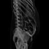
CT exams are not routinely ordered for pregnant women, but may be necessary to detect suspected life-threatening conditions such as bleeding in the brain, blood clots in the lungs or appendicitis. Since CT exposes the developing fetus to radiation, concerns are often raised regarding overuse. The majority of CT examinations (approximately 75 percent) analyzed in the study were performed in areas of the mother’s body separate from the uterus, so the fetus was not exposed to any direct radiation. Still, low levels of radiation have been shown to carry a small risk of harm to a developing fetus.
“Women should know that imaging is generally safe during pregnancy and is often used to detect potentially life-threatening problems,” Dr. Lazarus said. “However, this study should raise awareness about imaging trends in pregnant patients and help us continue in our efforts to minimize radiation exposure,” Dr. Lazarus said.
The researchers evaluated 5,270 examinations on 3,285 patients. During the 10 years of the study, the number of patients imaged per year increased from 237 to 449, and the number of exams per year increased from 331 to 732. This represented an 89 percent increase in patients and a 121 percent increase in examinations over the course of the study. During the same 10 years, the number of deliveries only increased 7 percent from 8,661 to 9,264. Imaging utilization rates (exams per 1,000 deliveries) increased 107 percent.
Use of plain-film x-rays increased an average of 6.8 percent per year, and the number of nuclear medicine examinations rose by approximately 11.6 percent annually. Fluoroscopy utilization increased by 10.6 percent per year, and CT examinations increased by 25.3 percent per year.
A milliGray (mGy) is a unit of measure for absorbed radiation. The average estimated fetal radiation exposure per exam for CT was 4.3 mGy, compared to 2.91 mGy for fluoroscopy, 0.40 mGy for nuclear medicine and 0.43 mGy for x-rays.
Dr. Lazarus hopes that increased use of electronic medical records will help physicians and patients keep track of the number and types of imaging tests performed on pregnant women and give proper consideration to alternative imaging tests - such as MRI and ultrasound - that do not expose the patient or fetus to ionizing radiation.
Source: “Utilization of Imaging in Pregnant Women: A Ten Year Review of 5270 Exams in 3285 Patients: 1997-2006.” Collaborating with Dr. Lazarus were Carolynn DeBenedectis, M.D., David North, Sc.M., Patricia K. Spencer, M.D., and William W. Mayo-Smith, M.D.
Radiology is edited by Herbert Y. Kressel, M.D., Harvard Medical School, Boston, Mass., and owned and published by the Radiological Society of North America, Inc. (RSNA.org/radiologyjnl)
For more information: www.rsna.org

 June 18, 2026


