June 24, 2016 — The British Heart Foundation (BHF) announced the winners of its annual ‘Reflections of Research’ image competition, reflecting the charity’s research into heart and circulatory disease.
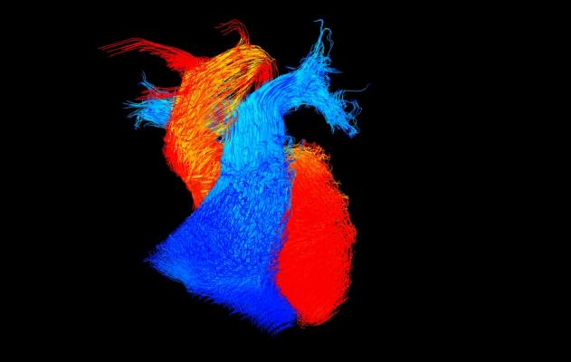
The winning image — titled “Go with the flow,” by Victoria Stoll, a BHF-funded researcher at the University of Oxford — captures the blood flowing within an adult heart frozen in time. Blood flows within the main pumping chambers (ventricles) of the heart and the vessels leaving the heart. The blue flow is blood that lacks oxygen and is travelling to the lungs. The red flow is blood that has been through the lungs and received oxygen and is now ready to be pumped around the body.
Stoll is using this type of imaging, four-dimensional cardiac magnetic resonance imaging (MRI), to look at the blood flow in four dimensions within the hearts of people with heart failure, whose hearts are not pumping effectively. She has already found that in people with severe heart failure the blood flows around the heart in a more disordered and disrupted pattern.
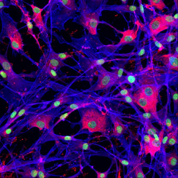
The Reflections of Research Supporters’ Favorite, chosen from an online vote on the BHF Facebook page, shows heart cells taken from newborns who received a surgical operation for correction of congenital heart defects shortly after birth. The blue color shows the shape of the cells, while the magenta shows a structural protein necessary to glue the cells together.
Dr. Elisa Avolio, a BHF-funded researcher at the University of Bristol, is using these cells to explore the possibility of treating congenital heart disease. Congenital heart defects are the most common type of birth defect, In the UK alone over 4,000 babies are diagnosed with congenital heart disease each year.
The only treatment for some of these conditions is corrective surgery where a piece of tissue, known as a graft, is used to replace the affected area. However, often surgery has to be repeated several times throughout childhood as the child’s heart outgrows the graft used to repair it.
Avolio is working on grafts that are able to grow like living tissue and can therefore grow along with a child’s heart. These new grafts would mean that instead of having multiple operations to insert bigger grafts as the patient’s heart grows, only one operation would be needed.
Prof. Peter Weissberg, medical director at the BHF and one of this year’s judges, said, “Science relies increasingly on ever more sophisticated imaging techniques to help us to see the cellular and molecular processes that conspire to create disease.
Each of these images contains a wealth of information that scientists can use in their fight against cardiovascular disease. So whilst this competition is all about stunning imagery, it’s actually the story that the image tells that matters.”
One of the judges of this year’s competition, wildlife photographer Andrew Rouse, said, “The winning image is simply beautiful. It’s both amazingly abstract and instantly recognizable. My 11-month-old daughter is fascinated by it, and she is perhaps the best judge of all showing that this image is simple yet also very striking, which is what a good photograph should be.”
Competition judge and artist Sophie Layton said, “Bringing the worlds of art and science together is such a perfect way to explore the wonders of science and the extraordinary insights that we are able to witness through the technology that scientists have access to. Finding an artistic expression can make these concepts accessible to a much wider community.”
For more information: www.bhf.org.uk

 July 10, 2026


