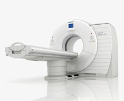
Stock image

March 9, 2009 - The use of dual-source computed tomography (DSCT) may be useful to noninvasively detect in-stent restenosis, especially in stents with a relatively large diameter, according to a study in this week’s issue of the American Journal of Cardiology (pages 812-817).

The study was conducted by researchers from the Department of Internal Medicine, University of Erlangen-Nuremberg, Erlangen, Germany, the Institute of Diagnostic Radiology, University of Erlangen-Nuremberg, Erlangen, Germany, and the Cardiac MR PET CT Program, Massachusetts General Hospital and Harvard Medical School, Boston. The assessed the accuracy of evaluating coronary artery stents using DSCT, because it provides higher temporal resolution that may allow more accurate evaluation of coronary stents. A total of 112 patients with 150 previously implanted coronary stents (diameter ?3 mm) were examined using a Definition DSCT made by Siemens Medical Solutions before conventional coronary angiography.

Each stent was classified as assessable or not assessable. All assessable stents were further classified for the absence or presence of in-stent restenosis (>50% diameter reduction) using DSCT, and results were compared with those using quantitative coronary angiography. Mean stent diameter was 3.27 ± 0.35 mm. Fifteen of 80 stents (19%) with a diameter of 3.0 mm were not assessable, and all 70 stents >3.0 mm were assessable. DSCT correctly identified 16 of 19 in-stent restenoses in 135 assessable stents, as well as the absence of in-stent restenosis in 110 of 116 stents (sensitivity 84%, specificity 95%, positive predictive value 73%, and negative predictive value 97% in assessable stents).

For more information: www.ajconline.org/article/S0002-9149%2808%2902091-2/abstract


Related Content

Feature | Computed Tomography (CT) | By Melinda Taschetta-Millane

Computed Tomography (CT) continues to be a rapidly evolving technology with many new advancements, as displayed and ...

Time March 07, 2024
arrow
News | Computed Tomography (CT)

February 20, 2024 — Ultrahigh-spatial-resolution photon-counting detector CT improved assessment of coronary artery ...

Time February 20, 2024
arrow
News | Contrast Media

August 2, 2022 – A new update has been announced as the radiology world continues to address supply disruptions of ...

Time August 02, 2022
arrow
News | Mobile C-Arms

July 29, 2022 — Siemens Healthineers has announced the Food and Drug Administration (FDA) clearance of the ARTIS icono ...

Time July 29, 2022
arrow
News | SCCT

July 8, 2022 — The Society of Cardiovascular Computed Tomography (SCCT) has released a new expert consensus document on ...

Time July 08, 2022
arrow
News | PET-CT

January 12, 2022 — By combining information from two advanced imaging techniques with clinical data, physicians can ...

Time January 12, 2022
arrow
Feature | Computed Tomography (CT) | By Dave Fornell, ITN Editor

The U.S. Food and Drug Administration (FDA) cleared the world's first photon-counting computed tomography (CT) scanner ...

Time November 04, 2021
arrow
News | Computed Tomography (CT)

October 14, 2021 — Cardiac computed tomography angiography (CTA) derived left atrium emptying fraction (LAEF) improves ...

Time October 14, 2021
arrow
News | Artificial Intelligence

September 27, 2021 — Zebra Medical Vision, the deep-learning medical imaging analytics company, announces its eighth U.S ...

Time September 27, 2021
arrow
News | Cardiac Imaging

September 22, 2021 — Test selection should be a shared decision between patient and physician rather than directed by ...

Time September 23, 2021
arrow
Subscribe Now