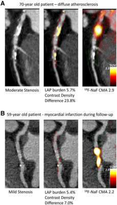
Case examples of quantitative plaque analysis on coronary CT angiography and 18F-NaF PET in patients with established coronary artery disease. Hybrid CT angiography and 18F-NaF PET of coronary arteries. (A) A 70-y-old male, who presented with diffused largely noncalcified disease (middle panel in red) in the LAD and demonstrated increased 18F-NaF uptake in the LAD on PET. (B) A 59-y-old male with mild LCX atherosclerosis, who presented with a high noncalcified plaque burden (middle panel in red) on CT angiography, significant 18F-NaF uptake and experienced a lateral non–ST-segment elevation myocardial infarction during follow-up. LAD = left anterior descending; LCX = left circumflex; LAP = low attenuation plaque. Image courtesy of first author Jacek Kwiecinski and senior author Piotr Slomka, Cedars-Sinai, Los Angeles, Calif., in collaboration with Edinburgh University, UK.
January 12, 2022 — By combining information from two advanced imaging techniques with clinical data, physicians can improve their prediction of heart attacks, according to research published in the January issue of The Journal of Nuclear Medicine. When assessed together in an artificial intelligence model, coronary 18F-NaF uptake on PET and quantitative coronary plaque characteristics on CT angiography were found to be complementary, strong predictors of heart attack risk in patients with established coronary artery disease, providing risk prediction superior to that of clinical data alone.
In everyday clinical practice, predicting a heart attack is challenging. The predicted likelihood of a heart attack typically is based on cardiovascular risk factors and scores, especially in patients with suspected coronary artery disease. However, in patients with confirmed coronary artery disease, cardiovascular risk factors and scores don’t always show the full picture.
“Recently, advanced imaging techniques have demonstrated considerable promise in determining which coronary artery disease patients are most at risk for a heart attack. These techniques include 18F-sodium fluoride (18F-NaF) PET, which assesses disease activity in the coronary arteries, and CT angiography, which provides a quantitative plaque analysis,” said Piotr J. Slomka, PhD, FACC, FASNC, FCCPM, director of Innovation in Imaging at Cedars-Sinai Medical Center in Los Angeles, Calif. “Our goal in the study was to investigate whether the information provided by 18F-NaF PET and CT angiography is complementary and could improve prediction of heart attacks with the use of artificial intelligence techniques.”
Nearly 300 patients with established coronary atherosclerosis participated in the study. All patients underwent a baseline clinical assessment with evaluation of their cardiovascular risk factor profile. All patients received hybrid coronary 18F-NaF PET and contrast CT coronary angiography. Machine learning—a type of artificial intelligence—was used to calculate a joint score for heart attack risk by incorporating key variables from the clinical assessment, 18F-NaF PET findings and quantitative CT variables.
The machine learning model showed substantial improvement in prediction of heart attack over clinical data alone. This approach demonstrated that 18F-NaF PET and CT angiography are complementary and additive, with the combination of both providing the most robust outcome prediction.
“18F-NaF PET combined with anatomical imaging provided by CT angiography has the potential to enable precision medicine by guiding the use of advanced therapeutic interventions,” noted Slomka. “Our study supports the use of artificial intelligence methods for integrating multimodality imaging and clinical data for robust prediction of heart attacks.”
For more information: www.snmmi.org
Related Multimodality Imaging Content:
Deep Learning with SPECT Accurately Predicts Major Adverse Cardiac Events
Using Contrast MRI After a Heart Attack Could Increase Survival
Medical Societies Support Safety and Benefits of Ultrasound Contrast Agents

 July 22, 2026


