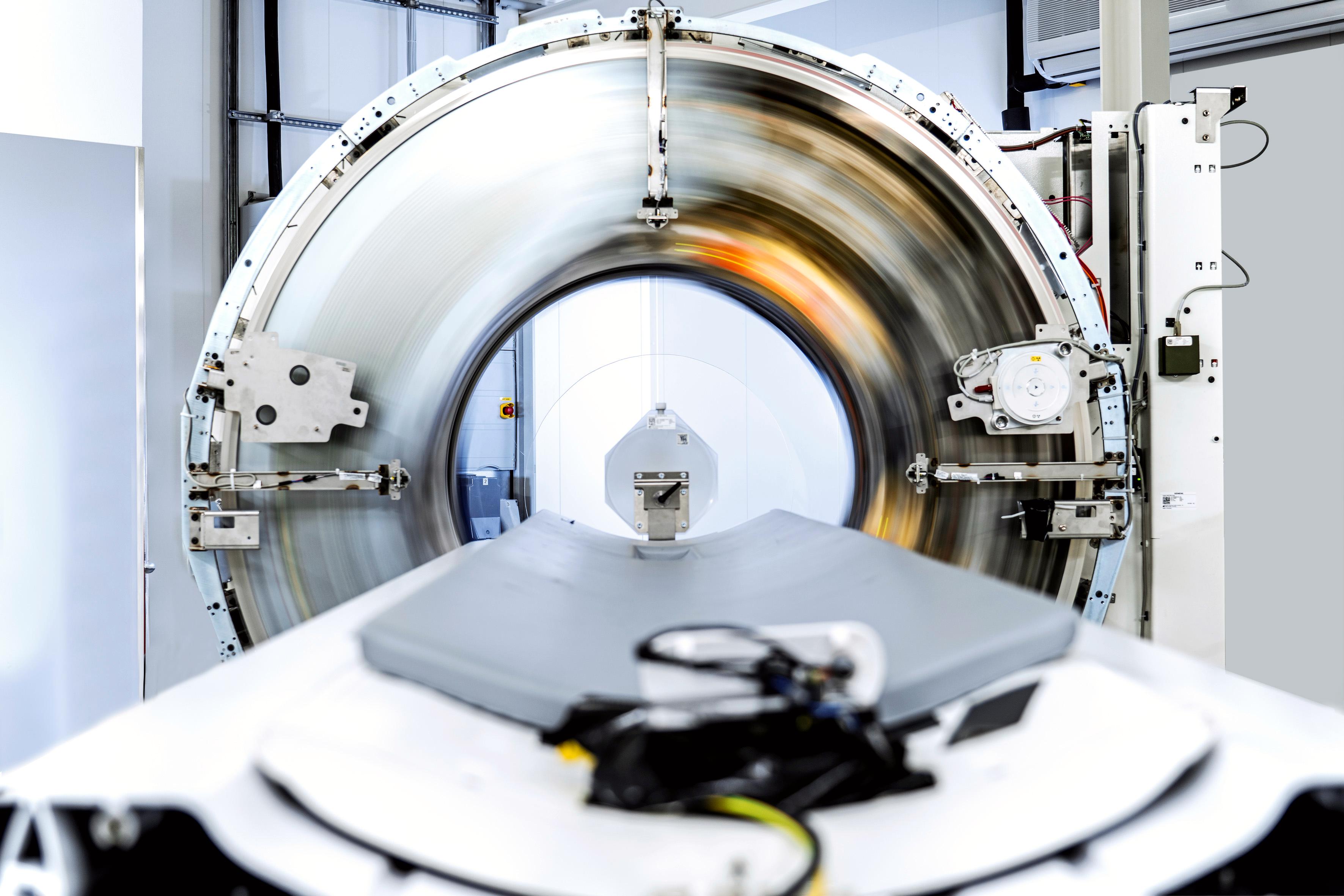
The Siemens Naeotom Alpha is the first commercialized photon-counting CT scanner. It gained FDA clearance Sept. 30, 2021. The image shows the system undergoing phantom testing with the outer covers off, showing the motion of the X-ray source and detector ring. Image by Deutscher Zukunftpreis/Ansgar Pudenz.
The U.S. Food and Drug Administration (FDA) cleared the world's first photon-counting computed tomography (CT) scanner, the Siemens Naeotom Alpha, on Sept. 30. Both FDA and CT experts say this is the start of a revolution in new CT scanner technology, and is the biggest shift in technology for this workhorse radiology modality in years.
The FDA sent out a rare press release on the approval, noting its importance to medical imaging.
“Computed tomography is an important medical imaging tool that can aid in diagnosing disease, trauma or abnormality; planning and guiding interventional or therapeutic procedures, and monitoring the effectiveness of certain therapies,” said Laurel Burk, Ph.D., assistant director of the Diagnostic X-ray Systems Team in the FDA’s Center for Devices and Radiological Health. “Today’s action represents the first major new technology for computed tomography imaging in nearly a decade and underscores the FDA’s efforts to encourage innovation in areas of scientific and diagnostic progress.”
The device uses the emerging CT technology of photon-counting detectors, which can measure each individual X-ray photon that passes through a patient's body, as opposed to current systems which use detectors that measure the total energy contained in many X-rays at once. By "counting" each individual X-ray photon, more detailed information about the patient can be obtained and used to create images with less information that is not useful, such as image noise.
The Siemens Naeotom Alpha is designed to transform the information from X-ray photons that pass through a patient's body, and are received by a detector, into a detailed 3-dimensional image.
"The use of photon-counting detectors in CT scanners is really a reinvention of CT imaging because the X-ray detector is the secret sauce that determines the quality of an image, somewhat like how the number of megapixels in your phone camera determines the quality of your photo," said Cynthia McCollough, Ph.D., the scientific director of the Mayo Clinic CT Clinical Innovation Center, which worked with Siemens in the development of the system and in testing system prototypes. She said photon-counting detectors are much better than the current generation of conventional CT scanners, and these properties will likely propel photon-counting detectors to became the next major shift in CT technology in the coming years.
"The images that we are seeing are incredible. We can appreciate structures that were simply too small to be resolved with previous detector technologies," McCollough said. "The resolution is exquisite, there are noise benefits, and it is dual-energy plus, because you can get multiple energies from one scan."
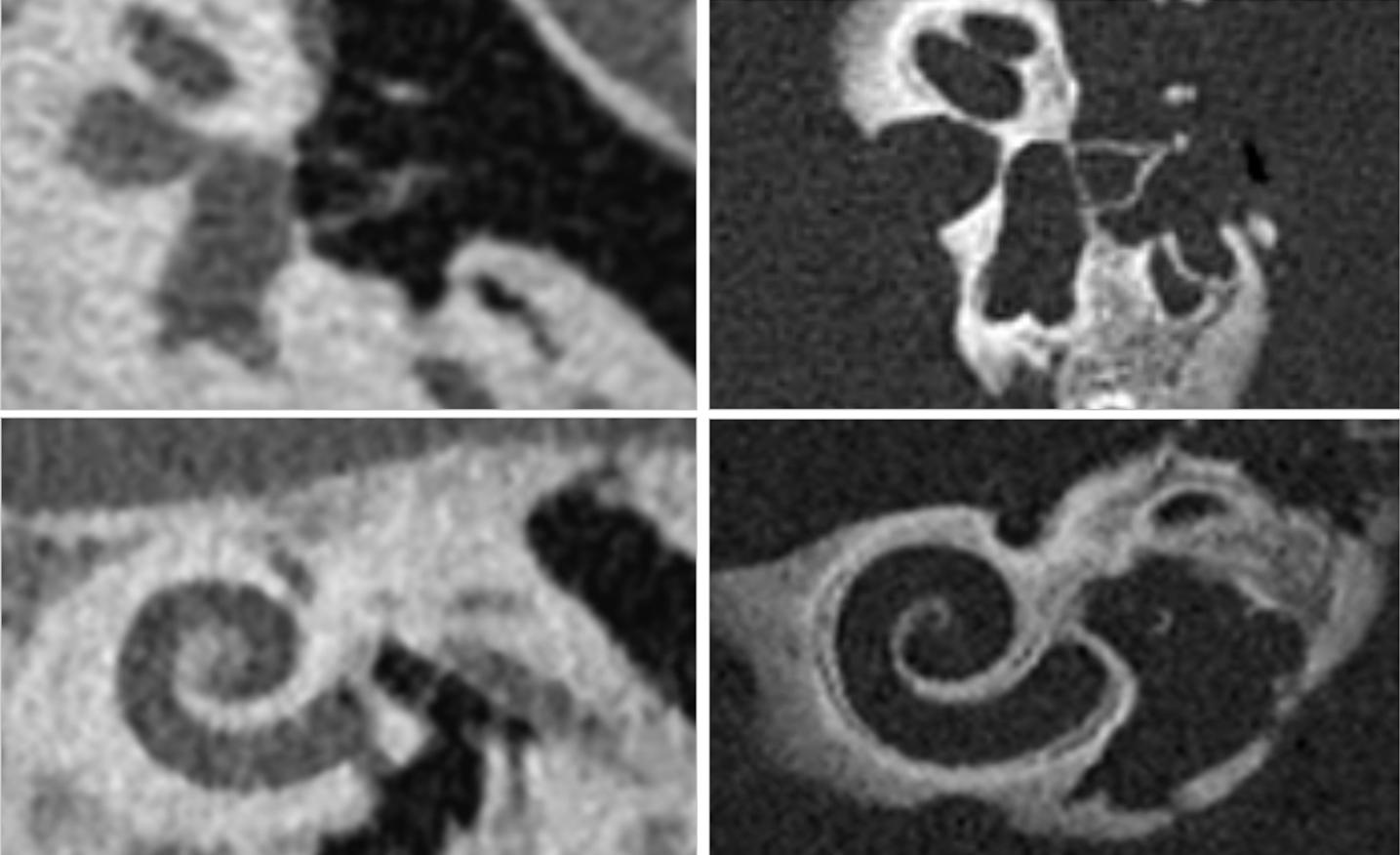
Improved visualization of the middle and inner ear enabled by photon-counting CT. Tiny anatomical structure of the middle and inner ear such as the stapes (upper row), and the cochlea (lower row). The left images were acquired using conventional CT imaging and the images on the right use photo-counting CT. Images courtesy of Dr. A. Persson, Linkoping University, Sweden.
New Type of CT Detector Improves Image Quality, is More Efficient and Collects More Data
The centerpiece of the Naeotom Alpha innovation is the new photon-counting detector, which uses an active detection layer that offers advantages over conventional CT detectors. Current CT technology uses a two-step conversion process to convert X-ray photons into visible light using a scintillator layer in the detector. Then, photo diode light sensors turn the visible light into a digital signal. Due to this intermediate step, important information about the energy of the X-rays is lost and no longer available to aid in diagnosis. Also, contrast is reduced and images are not as clear.
Photon-counting detectors use a single step of direct conversion of X-rays into electrical current, and skips the step of converting X-rays into visible light. This allows the energy thresholds of each pulse to be collected and binned based on different kilovolt (kV) energy levels. This creates data to improve contrast and enable dual-energy, spectral imaging. The direct conversion also helps improve image quality without information loss. This improves image sharpness and contrast.
Photon-counting detectors have already been used for several years in high-energy physics and nuclear imaging. However, these previously generation photon-counting detectors could not be used with a clinical CT scanner because they could not keep up with the high higher rate of photons reaching the detector. The detector on the Naeotom Alpha was designed for this increased speed.

Close-up of the photon-counting sensor boards inside the Naeotom Alpha CT scanner. Image by Deutscher Zukunftpreis/Ansgar Pudenz.
What Are the Benefits of Photon-counting CT:
Experts in CT say the benefits of photon counting systems include the following:
• No electric noise: This leads to better image quality in obese patients and in low-dose scans.
• Smaller detector pixels: This will greatly improve spatial resolution to improve image quality over conventional CT scanners and help further reduce noise.
• No down-weighting of lower energy photons: This improves image contrast, including iodine contrast to noise ratio (CNR).
• Intrinsic spectral imaging built into each scan: All photon-counting scans have a spectral CT component because the detector bins photons of different energies, allowing them to be accessed in post-processing to show the image at other energies. These scanners will eliminate the need for dedicated dual-energy CT scanners.
• Direct digital detection of photons without a scintillator: Removing the two-step conversion process to convert X-ray photons into visible light first helps improve image quality. This also allows photon-counting detectors to differentiate the energy of each photon to enable dual-energy, spectral imaging.
"This is really a change at the detector level with a fundamental change in how the photons emitted by the CT scanner are — as the name says — counted," explained Todd Villines, M.D., FACC, FAHA, MSCCT, is the Julian Ruffin Beckwith Professor of Medicine, Division of Cardiovascular Medicine, University of Virginia, editor-in-chief of the Journal of Cardiovascular CT (JCCT), and past-president or the Society of Cardiovascular Computed Tomography (SCCT). "It is a much more efficient way to use photon energy, so not only is it lower dose, but the image quality, particularly related to stents and calcium, appear to be much better than standard detectors."
The Value of Spectral Imaging Incorporated Into Every Photon-counting CT Scan
The spectral properties of photon-counting will enable radiologists or cardiologists to peel back metal or calcium on a molecular level in scans to reveal the tissue or plaque build up inside stents or in vessels packed with calcium deposits, Villines explained. The chemical decomposition ability of spectral imaging also can help reduce metal artifact blooming in images, not only for stents or prosthetic valves, but also for orthopedic implants and surgical staples that can often obscure the view in images.
These new detectors can take the bins of photons collected at various energies to reconstruct a range of mono-energtic image renderings similar to dual-energy CT, but on a wider spectrum of kV levels. This spectral aspect of photon counting also allows material decomposition based on the chemical elements that make up various materials in the scan, including iodine contrast, calcium and metals that make up stents, orthopedic implants and replacement heart valves. This enables easier, automated removal of metal blooming artifacts and the ability to clearly image inside calcified arteries.
"We will be able to be more confident in scanning patients who have stents or who have highly calcified coronary arteries," Villines said. "The implications of photon-counting CT are really profound. The beauty of photon-counting detectors is that it allows for direct conversion of emitted X-rays into energy, and this is something that is much more efficient. It also allows you to bin different energies across a variety of mono-energenic kVs. It also has increased spacial resolution, reduced noise and uses lower radiation dose."
He said another advantage to the spectral abilities of photon counting is the ability to separate various contrast agents, such as iodine, gadolinium and bismuth, so multi-contrast types can be injected at the same time and the imaging system can show each individual contrast type image, or combine them for much higher soft tissue resolution that is closer to MRI. He said research is already being done on this and he suspects multi-contrast agnate scanning may because a trend in the next decade as photon-counting scanners are commercialized and put into service.
"MRI has always had really good tissue characterization, and I think CT has the promise to do that as well with this new technology," Villines said.
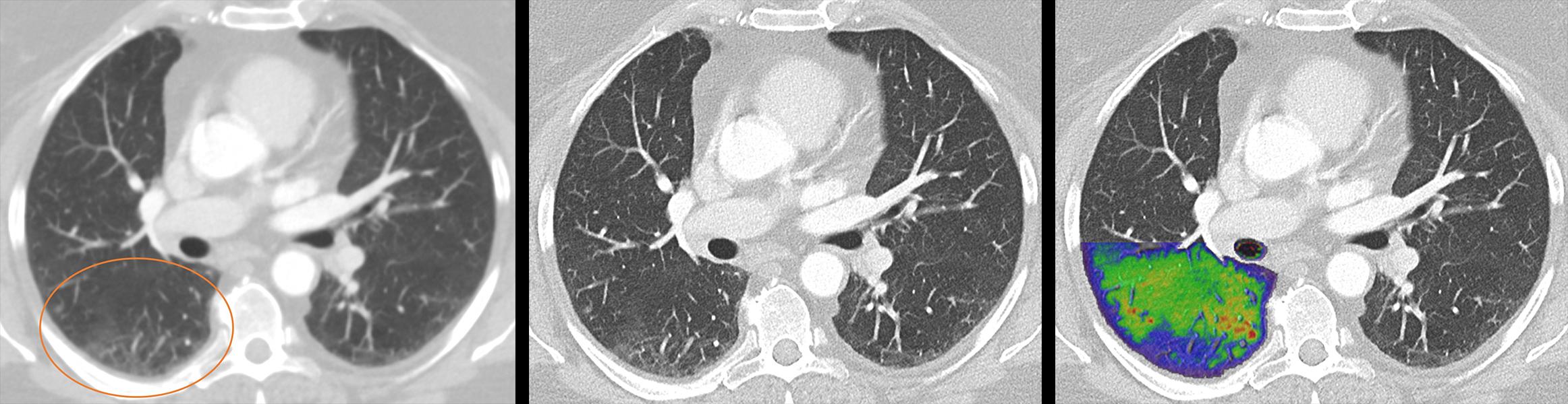
Lung images of a post-COVID patient with photon-counting CT. The photon-counting technology allows simultaneous acquisition and visualization of detailed structures (center image) combined with functional information (right image). For comparison is an image with conventional CT (left). Courtesy of Dr. J. Ferda, University Hospital Plzen, Czech Republic.
New Scanner Has Advantages in Cardiac Imaging
The Siemens Naeotom Alpha also uses a unique geometry to perform state-of-the-art cardiac imaging. In order to get clear, crisp images of the heart, which is always moving, the system needs to be fast.
"And this system is extremely fast," McCollough said. "Cardiac imaging is like taking a photograph of a bicycle rider going by and wanting to count the number of spokes in the wheel, but the spokes are blurred because they were moving. With the new system, we can capture a small fraction of one heartbeat, which freezes the motion."
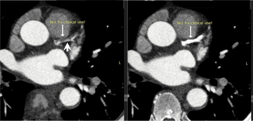
The inherent spectral capabilities of Mayo Clinic's new third-generation photon-counting CT research scanner allows clear visualization of the intact portions of the coronary artery lumen (arrowhead) by reducing the bright signal from the dense coronary calcifications (arrows). Spectral CT is able to bin photos on different energies that can be used to enhance or subtract chemical elements from the images based on their spectral signature, such as calcium shown here, and iodine.
Development of Photon-Counting CT Systems
The CT Clinical Innovation Center at Mayo Clinic in Rochester, Minn., has been involved for almost a decade in discovering how the emerging technology of photon-counting detector CT might benefit patients. The work is part of a collaboration with Siemens Healthineers, which developed the prototype system.
Mayo Clinic in Rochester installed the world's first photon-counting-detector CT system capable of human imaging in 2014. The first research studies in humans began in August 2015. Since then, the center has worked closely with the manufacturer to evaluate the clinical potential of the technology.
A second-generation prototype debuted at Mayo in 2020. This system addressed many of the design limitations of the first system. The third-generation research system has been up and running since April 2021 as part of the ongoing Siemens research collaboration. In April, Mayo Clinic performed its first cardiac scan on the new system. The completion of that exam elicited a round of applause from the research team.
"This type of advance in an imaging modality only comes along every couple of decades," said Joel Fletcher, M.D., medical director of the Mayo CT Clinical Innovation Center. He said previous major CT technology innovations included spiral CT, multidetector-row CT and dual-energy CT. Fletcher predicts that over the next five or 10 years, photon-counting-detector CT will lead to major advances in medical imaging that will result in improved patient care.
"It's a special time in all of our careers when we have these paradigm-shifting technical advances," Fletcher said.
Siemens also said the collaboration with Mayo and other hospitals on this project has been a major success.
“About 15 years ago, work on photon counting and its clinical vision started at Siemens Healthineers. We always believed in the tremendous clinical value and relentlessly worked on it together with our partners. We are excited that we have received FDA 510(k) clearance,” said André Hartung, head of diagnostic imaging at Siemens Healthineers.
Photo-counting CT Systems Are Being Developed by All Major CT Vendors
All major imaging companies are developing photon-counting CT detectors. While this technology has been researched academically for several years, it appears several vendors are close to commercializing these detectors, with Siemens only being the first to market.
At RSNA 2020, GE Healthcare said photon-counting CT detector technology is the way of the future and highlighted its purchase of the Swedish company Prismatic Sensors AB in November 2020. The company is developing photon-counting, deep silicon detectors that GE will use for its prototype photon counting CT system. GE began operation of this prototype system in 2021.
Samsung is working with a large U.S. university to develop its PCD technology and discussed the new detector at RSNA 2020.
Philips Healthcare also has prototype scanners in operation.
Related Photon-counting CT Content:
Mayo Clinic Begins Use of Third-Generation Photon-counting CT Clinical Research Detector
VIDEO: New Advances in CT Imaging Technology — Interview with Cynthia McCollough, Ph.D.
VIDEO: Photon Counting Detectors Will be the Next Major Advance in Computed Tomography — Interview with Todd Villines, M.D.
Key Trends in Cardiac CT at SCCT 2020
GE Healthcare Pioneers Photon Counting CT with Prismatic Sensors Acquisition
Top Trend Takeaways in Radiology From RSNA 2020
VIDEO: Advances in Cardiac CT Imaging — Interview with David Bluemke, M.D.


 July 14, 2026


