
Feature
As health IT leaders make their way to New York for the New York eHealth Collaborative (NYeC) Digital Health Conference, Practice Fusion, the nation's largest healthcare platform, is looking at the key healthcare trend from 2013 and predicts what they might mean looking forward.
 November 14, 2013
News
Accuray Inc. announced the publication of two papers stemming from a large multi-center study of CyberKnife stereotactic body radiotherapy (SBRT) led by investigators at the University of California, Los Angeles (UCLA).
November 14, 2013 News
Royal Philips Electronics and Sectra announced an agreement to extend the term of their existing picture archiving and communication system (PACS) support partnership agreement.
November 14, 2013 Sponsored Content
Sponsored Content
Videos | Radiology Imaging
Today’s radiology teams are faced with a wide range of growing complexities such as patient and procedure variabilities ...
June 24, 2026 
Technology
DICOM Grid, a provider of cloud-based medical image exchange solutions, announced the world-wide availability of DG Revenue Engine, a service designed to turbo-charge patient portals for second opinions.
November 14, 2013 
Technology
Clear Image Devices LLC (CiD) announced the release of its step platform to be used for standing patient ultrasound venous insufficiency studies. The Ultrasound Exam Steps enable sonographers to place the patient in the optimal position for safe, easy capture of the highest quality venous images of the upper and lower leg.
November 14, 2013 News
The Chinese market for X-ray equipment will grow 50 percent from 2012 levels to reach more than $1.5 billion by 2017, according to a new report from HIS.
November 14, 2013 Sponsored Content

Feature | Information Technology
AT A GLANCE Organization: Expert Radiology Management Services, LLC Specialty: Subspecialty teleradiology — neuro and ...
May 01, 2026 News
To help reduce the burden of cardiovascular disease, the nation's leading killer, New York-Presbyterian Hospital and Weill Cornell Medical College have created the Dalio Institute of Cardiovascular Imaging. Raymond T. Dalio, a life trustee of New York-Presbyterian Hospital, has made a gift of $20 million through his Dalio Foundation in support of the institute.
November 14, 2013 
News
Picture Archiving and Communication System (PACS) vendor DR Systems will exhibit new capabilities designed to increase the quality and efficiency of patient services at the Radiological Society of North American Annual Meeting (RSNA) 2013.
November 14, 2013 Feature
The Eurozone economic slowdown has made it difficult for hospitals, particularly in Southern Europe, to procure new imaging modalities. With streamlined budgets and escalating number of medical procedures, the need to spend less on technologies while gaining the maximum benefit from them has sustained demand for high-quality refurbished imaging equipment.
November 14, 2013 Sponsored Content
Sponsored Content
Videos | Radiology Business
Radiology departments have many different needs and face a wide variety of challenges that can impact their departments ...
November 11, 2025 
Technology
Radcal Corp. will launch its new models of diagnostic X-ray meters and sensors, Accu-Gold+ and Rapid-Gold+ with improved stacked sensor multisensors including a dual rad/fluoro and mammo all-in-one multisensor, at the Radiological Society of North American Annual Meeting (RSNA 2013).
November 14, 2013 
News
Indianapolis-based Modular Devices Inc. reported it expanded its fleet of interim labs to include more computed tomography (CT) scanner options. The turnkey labs are available to medical centers around the country for short and long-term leases.
November 14, 2013 News
A University of Colorado Cancer Center study, published in the journal Physics in Medicine and Biology, shows that endorectal balloons commonly used during precise radiation treatment for prostate cancer can deform the prostate in a way that could make radiation miss its mark.
November 14, 2013 Sponsored Content
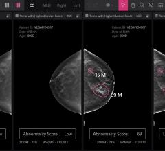
Feature | Breast Imaging
Despite decades of progress in breast imaging, one challenge continues to test even the most skilled radiologists ...
October 24, 2025 
News
Lantheus Medical Imaging, Inc., a developer, manufacturer and distributor of diagnostic imaging agents, announced preliminary results from the first of two planned Phase 3 trials to assess the diagnostic efficacy of flurpiridaz F 18, an imaging agent used in positron emission tomography (PET) myocardial perfusion imaging (MPI) for the detection of coronary artery disease (CAD).
November 13, 2013 
Feature
No law prevents doctors from freely prescribing U.S. Food and Drug Administration (FDA)-approved drugs and devices for off-label uses, yet regulators continue to aggressively pursue civil and criminal enforcement of perceived violations, warns Patrick J. Hurd, senior counsel with LeClairRyan in Washington, D.C. and Norfolk, Va. in the November 2013 edition of Westlaw Journal Pharmaceutical.
November 13, 2013 News
Virtual Radiologic (vRad), a radiology practice, announced the release of its radiology patient care (RPC) Indices, the first set of findings-based national and peer group radiology benchmarking metrics.
November 13, 2013 
Technology
Intelerad Medical Systems announced the launch of InteleConnect Patterns, an optional service built into the InteleConnect Referring Physician Portal that provides radiology groups with key analytics regarding their base of referring physicians.
November 12, 2013 
Technology
Agfa HealthCare announced that it is launching the next generation of its MUSICA (Multi-Scale Image Contrast Amplification) image processing software with new technology improvements that enhance both image quality and workflow for radiographers and radiologists at the upcoming Radiological Society of North America Annual Meeting (RSNA 2013) in Chicago.
November 12, 2013 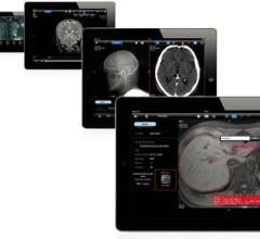
Technology
Aycan will feature its mobile iPad application, which is designed to quickly, easily and securely transfer DICOM images from hospitals and imaging centers to on-call and other radiologists and referring physicians with an iPad, at the Radiological Society of North America Annual Meeting (RSNA 2013) in Chicago.
November 12, 2013 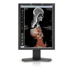
Technology
NEC Display Solutions will be offering additional insight and demonstrations of its latest products including the MultiSync MD210C2 medical-grade monitor, built for the displaying and viewing of digital images for diagnosis by physicians, at the Radiological Society of North America Annual Conference (RSNA 2013) in Chicago.
November 12, 2013 
Feature
Personnel of Wyle, a provider of engineering, scientific and technical service to the U.S. Department of Defense (DoD), at the Johnson Space Center have selected Fujifilm Medical Systems U.S.A. Inc. to implement Synapse Radiology and Synapse Cardiovascular to support NASA’s in-flight as well as ground-based clinical care operations.
November 12, 2013 © Copyright Wainscot Media. All Rights Reserved.
Subscribe Now