Varian Medical Systems launched RapidPlan Knowledge-based Planning at ASTRO 2013. RapidPlan is a new paradigm for ...
Through their network of hospitals, clinics, centers, health units, and residential facilities, the Island Health (Vancouver Island Health Authority) provides healthcare to more than 765,000 people located in a geographic area of approximately 34,800 square miles.Spanning across nineteen medical imaging sites on Vancouver Island, Island Health boasts a highly skilled team of professionals who work together to provide diagnostics and treatment procedures.An Intelerad partner for over 15 years, Island Health uses IntelePACS and InteleViewer to read over 800,000 studies per year across Island health’s network of hospitals. “By leveraging Intelerad solutions, we have been able to greatly improve our medical imaging department’s efficiency,” said Ken Situ, a technical analyst with Island Health. “In particular, it has greatly reduced turnaround time, which allows us to provide reports to referring physicians quickly.”
ContextVision released another ultrasound product for the mid-end ultrasound segment; a new version of the image enhancement software US PlusView was designed for the digital signal processor (DSP).
Radiology departments have many different needs and face a wide variety of challenges that can impact their departments ...
International Business Machine (IBM) Research unveiled two new Watson-related cognitive technologies that are expected to help physicians make more informed and accurate decisions faster and to cull new insights from electronic medical records (EMR).
Digisonics Inc. introduced functionality for appropriate use criteria (AUC) calculations. Digisonics recognizes that reimbursement audits will be tied to appropriate use scores in the future, benefitting facilities that utilize the Digisonics CVIS (cardiovascular imaging and information systems) to monitor appropriate use scores and produce the required structured reports.
Voices Against Brain Cancer (VBAC), an organization dedicated to brain cancer research and advocacy, discussed a study that looked into the preservation of cognitive function for brain cancer patients after radiotherapy treatments.
Despite decades of progress in breast imaging, one challenge continues to test even the most skilled radiologists ...
Samsung Medison announced it is showcasing two new ultrasound systems including the UGEO WS80A, the first ultrasound device for obstetrics and gynecology (OB/GYN) at the 23rd International Society of Ultrasound in Obstetrics and Gynecology (ISUOG) World Congress.
DePuy Synthes CMF (craniomaxillofacial) announced the launch of Trumatch CMF Solutions for facial reconstruction, orthognathic surgery, distraction and cranial reconstruction at the 2013 American Association of Oral and Maxillofacial Surgeons (AAOMS) annual meeting.
Departmental picture archiving and communication system (PACS) dominates the overall vendor neutral archive (VNA) market and PACS market with around 86.5 percent of the total revenue contribution, generating most of the revenues from PACS replacements. Hence, the market is expected to witness a stable compound annual growth rate (CAGR) of 5.2 percent from 2013 to 2018. Enterprise PACS, on the other hand, is growing faster than departmental PACS and is expected to reach $510 million by 2018 at an almost double CAGR.
Bayer Radiology’s Barbara Ruhland and Thom Kinst discuss how radiology departments can address the many different ...
DePuy Synthes Spine announced it is expanding its collaboration with Brainlab through the worldwide launch of navigation-ready instrumentation for its spine systems and an exclusive global agreement to co-market Airo Mobile Intraoperative CT (computed tomography) by Brainlab.
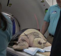
Oyster, a 26-year-old sea turtle, had an abnormal left flipper and shoulder for her whole life. There seemed to be a progressive muscle atrophy and disuse of the flipper, so Sea Life Minnesota sent her to the University of Minnesota Veterinary Medical Center (VMC) to do a computerized tomography (CT) scan on her left flipper and shoulder to learn more about the cause.
The 99th Scientific Assembly and 2013 Annual Meeting of the Radiological Society of North America (RNSA) will host more than 50,000 attendees from around the world and will feature a special lecture by former U.S. Secretary of State Condoleezza Rice, Ph.D., at 1:30 p.m. on December 3.
eHealth Saskatchewan plays a vital role in providing IT services to patients, health care providers, and partners such ...
To recognize the advances in the field of nuclear and molecular imaging, as well as the professionals who carry out these procedures, the Society of Nuclear Medicine and Molecular Imaging (SNMMI) and the SNMMI Technologist Section (SNMMI-TS) celebrated Nuclear Medicine and Molecular Imaging Week, October 6-12, 2013. The theme of this year’s Nuclear Medicine and Molecular Imaging Week was “Molecular Imaging: The Future…Delivered.”
People over 60 years old are more likely to have precancerous or cancerous polyps develop in a part of the colon that goes unseen by flexible sigmoidoscopy, a common screening test for colon and rectal cancer, a new study finds. Study results suggest the need for reviewing and possibly revising national colon cancer screening guidelines, study authors said at the 2013 Clinical Congress of the American College of Surgeons.
Breast cancer experts at Seattle Cancer Care Alliance (SCCA) developed guidelines to simplify recommendations from the American Cancer Society (ACS) on breast cancer prevention and screening.
High levels of high-density lipoprotein (HDL) have been linked to increased breast cancer risks and enhanced cancer aggressiveness in animal experiments.
Medical evidence over the past decade has demonstrated that patients with terminal cancer who receive a single session of radiotherapy get just as much pain relief as those who receive multiple treatments. But despite its obvious advantages for patient comfort and convenience (and cost savings), this single-fraction treatment has yet to be adopted in routine practice, according to a study led by the Perelman School of Medicine at the University of Pennsylvania published in The Journal of the American Medical Association (JAMA).
Olympus announced the launch of its U.S. Food and Drug Administration (F.D.A.)-cleared and world's only forward-viewing curvilinear ultrasound gastrovideoscope.
The Rotary Club of Park Ridge is making available its library, used to build and equip medical radiology facilities in clinics and small hospitals located in developing nations, free of charge on its website. The information provided by the library is practical and pragmatic, based on experiences of designing, equipping and installing X-ray rooms in Africa, Central Europe, Latin America and the Caribbean.
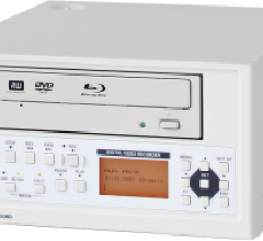
MAM-A Inc., a recordable discs supplier, announced its partnership with TEAC Corp. for the sales of the UR-50BD High Definition (HD) Medical Image Recorder. The medical image recorder is available together with medical-grade recordable DVDs, Blu-ray discs, or USB flash drives and is ideal for radiology, angiography, fluoroscopy, endoscopy and ultrasound. Discs and flash drives can have hospital logos imprinted on the surfaces if requested.


 October 16, 2013

