Researchers at the University of California Irvine recent published their findings on a new technology that achieves 3D imaging with a single X-ray projection called X-ray–Induced Acoustic Computed Tomography.
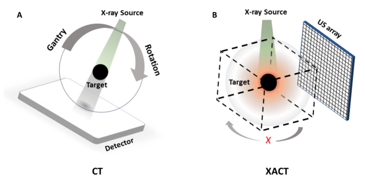
Computed tomography (CT) has long been a cornerstone of modern imaging, providing detailed 3D insights into the human body and other materials. However, conventional CT requires hundreds of X-ray projections from multiple angles, exposing patients to significant radiation doses and relying on large, immobile systems. To address this issue, researchers from UC Irvine's Departments of Radiological Sciences and Biomedical Engineering recently published a study in the journal Science Advances in which they introduce a groundbreaking technology that achieves 3D imaging with a single X-ray projection called X-ray–Induced Acoustic Computed Tomography (XACT).
A New Paradigm in Imaging
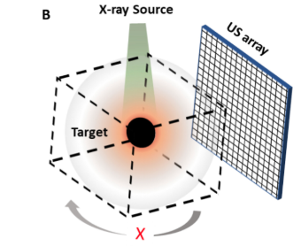
"In XACT, the generated sound waves by X-rays change the way X-ray imaging works, converting X-rays to ultrasound. X-rays typically travel in straight lines, so one projection only provides 2D information. However, X-ray-induced acoustic signals propagate in three dimensions, allowing for 3D imaging with a single projection," said Shawn Xiang, PhD, the study’s corresponding author and an associate professor at UCI’s Departments of Radiological Sciences and Biomedical Engineering.
XACT leverages the interaction between X-rays and tissue to produce acoustic waves, which travel at a speed of 1,500 meters per second. These waves are captured by ultrasound detectors, enabling real-time, three-dimensional imaging without the need for mechanical scanning or complex gantry systems.
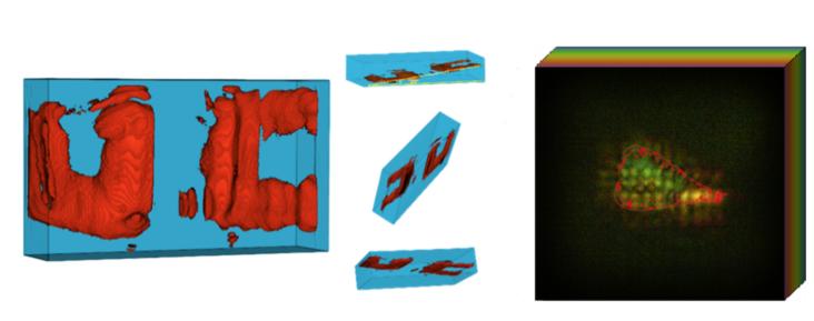
"For the first time, we have proved that 3D imaging can be obtained with a single X-ray projection based on X-ray-induced acoustic detection in both phantoms and biological tissue," said Siqi Wang, PhD, the study’s first author. Wang completed his PhD at UCI in Xiang’s lab and is now a postdoctoral research scholar at Stanford University.
"The groundbreaking finding here is that you can make 3D X-ray imaging with just a single projection, which typically needs 600 projections or more," says Vahid Yaghmai, MD, MS, FSAR, a radiologist at UC Irvine and chair of the UC Irvine Department of Radiological Sciences, who was not directly involved in the study.
Benefits Beyond Traditional CT
One of XACT’s most significant advantages is its efficiency and reduced radiation exposure. This makes XACT a safer and more accessible alternative, particularly for applications like routine diagnostics and breast cancer screening. Furthermore, with portable X-ray sources and ultrasound detectors, XACT systems promise compact, gantry-free designs, enabling imaging in settings previously inaccessible to traditional CT systems.
Challenges and Future Directions
While the potential of XACT is immense, current limitations include resolution constraints tied to the frequency and size of the ultrasound detectors. Future improvements, such as higher-frequency transducers and advanced reconstruction algorithms powered by deep learning, could further enhance its performance.
Redefining Imaging Across Fields
The ability to achieve 3D imaging from a single X-ray projection positions XACT as a transformative tool not only for medical diagnostics but also for nondestructive testing in engineering and material science. Its innovative approach eliminates the need for rotational access, opening new possibilities for imaging in constrained environments.
XACT also represents a leap forward in imaging technology, combining reduced radiation exposure, compact system design and unprecedented efficiency. As this technology continues to evolve, it has the potential to redefine medical and industrial imaging, bringing us closer to a future where high-resolution, low-dose 3D imaging is the norm in healthcare and beyond.

June 15, 2026 — Lead Glass Pro, a supplier of radiation shielding products, has expanded its turnkey installation ...
 June 18, 2026
June 1, 2026 — Serac Healthcare Ltd. has presented Phase 2 data showing that SPECT-CT imaging with the radiotracer 99mTc ...
June 01, 2026 
May 18, 2026 — DICO, a company specializing in the creation of distributed diagnostic infrastructure for radiology, has ...
May 19, 2026 
April 29, 2026 — Results from a new study* presented at the American Roentgen Ray Society’s (ARRS) 2026 annual meeting ...
April 29, 2026 
April 28, 2026 — Avatar Medical has been granted FDA 510(k) clearance for Avatar Medical Vision, its software platform ...
April 28, 2026 April 28, 2026 — Abbott has received U.S. Food and Drug Administration (FDA) clearance and CE Mark for its next ...
April 28, 2026 
April 16, 2026 — Royal Philips has received U.S. Food and Drug Administration 510(k) clearance for the Philips Spectral ...
April 20, 2026 
April 7, 2026 — Onvida Health and Siemens Healthineers have entered a 10-year Value Partnership¹ designed to bring the ...
April 09, 2026 
March 31, 2026 — Radon Medical Imaging, a medical imaging equipment maintenance and repair services company, has has ...
March 31, 2026 
March 26, 2026 — GE HealthCare has announced a renewed research collaboration with Stanford Medicine Department of ...
March 30, 2026