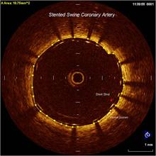
An optical coherence tomography (OCT) image of a stent in a coronary vessel. The shadows are from the stent struts.
May 5, 2010 – A new high-definition intravascular imaging system gained U.S. Food and Drug Administration (FDA) clearance today. LightLab Imaging Inc. said its C7-XR optical coherence tomography (OCT) imaging system and companion C7 Dragonfly OCT imaging catheter are now available on the U.S. market.
OCT offers near photographic quality imaging inside vessels and is a competing technology with intravascular ultrasound (IVUS). OCT is already available in more than 35 countries in Europe and Asia. The FDA clearance marks the first OCT intravascular imaging system approved for use in the United States.
The imaging system and catheter incorporate LightLab's Frequency Domain OCT (FD-OCT) technology. The C7-XR imaging system and the monorail style C7 Dragonfly catheter create a high-resolution 50 millimeter coronary scan in less than three seconds without the vessel occlusion that was required by earlier generation OCT systems. Thousands of data points are analyzed simultaneously at high speeds, providing intravascular resolution at 15 micrometers, roughly twice the size of a red blood cell. This nonocclusive, intravascular imaging technology allows the clinician to see and measure important vessel characteristics that are otherwise difficult to visualize with IVUS or angiography.
"The C7-XR Imaging System represents a significant step forward for intracoronary imaging," said Ik-Kyung Jang, M.D., Ph.D., professor of medicine at Harvard Medical School, director, cardiology laboratory for integrative physiology and imaging, Massachusetts General Hospital, Boston. He served as principal investigator for LightLab's U.S. clinical study. "We were all amazed with the speed and simplicity of the LightLab OCT procedure, and the clinical utility is truly unique. I expect OCT to rapidly become the new intracoronary imaging standard."
"LightLab's C7-XR FD-OCT image resolution is extraordinary, but what is most fascinating about this technology is its ease of use and extremely fast image acquisition," said Marco Costa, M.D., Ph.D., professor of medicine, director, interventional cardiovascular center and director, Center for Research and Innovation, Harrington-McLaughlin Heart and Vascular Institute University Hospitals, Case Western Reserve University. "These features will enable optimization of drug-eluting stent procedures with unprecedented accuracy, potentially reducing the number of stents placed per patient by ensuring appropriate disease assessment and targeting. The physician armed with OCT will have the ability to see the lumen vividly from the inside giving us incredible information and the ability to treat suboptimal results while the patient is still in the cath lab."
LightLab Imaging will demonstrate the C7-XR imaging system and C7 Dragonfly catheter May 5 to 8 at the Society for Cardiovascular Angiography and Interventions (SCAI) 2010 Scientific Sessions in San Diego, in booth 28.
LightLab's U.S. clinical study of the C7-XR imaging system and C7 Dragonfly catheter was conducted at three U.S. centers. The centers and the investigators were: Massachusetts General Hospital: Ik-Kyung Jang, M.D., Ph.D.; Columbia University Presbyterian Hospital: Jeffrey Moses, M.D., FACC, Giora Weisz, M.D., George Dangas M.D., Ph.D., and Varinder Singh, M.D.; and Stanford University Medical Center: William Fearon, M.D. and Alan Yeung, M.D. The core laboratory responsible for analyzing the study results was the University Hospitals Case Medical Center-Interventional Cardiovascular Center and Research and Innovation Center: Marco Costa M.D., Ph.D. and Hiram G. Bezerra, M.D., Ph.D.
For more information: www.lightlabimaging.com

 June 18, 2026