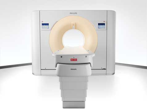
Philips Healthcares Brilliance iCT, a 256-slice CT, images the heart within two heartbeats with 80 percent less dose.
While medical imaging is considered one of the greatest technological advances in the last century, it does not come without risks. There has been growing debate amongst physicians, manufacturers and the medical community as a whole regarding patient exposure to radiation.
Although the exact amount of radiation exposure that is unsafe is not clearly defined, the U.S. Food and Drug Administration acknowledges: “The principal risk associated with the radiation dose resulting to a person from a CT procedure is the small possibility of developing a radiation-induced cancer some time later in that person’s life.”1 However, the FDA’s position is that the risk to benefit ratio depends on the patient’s condition.
Specifically in cardiac imaging, the widespread use of computed tomography (CT), angiography, and single photon emission computed tomography (SPECT) for exams has prompted clinicians to take action toward lowering radiation dose during exams. The trade off in CT is that, in most cases, the more radiation that is emitted, the more anatomical data is acquired. However, with the introduction of the highest powered CT system, a 320-slice volumetric CT scanner, it is so fast, it results in less radiation exposure. “Many people might think that Toshiba’s 320-slice scanner produces excessive radiation, compared to a 64-slice machine. But, in fact, the opposite is true because it is able to scan an entire heart in one gantry rotation of 300 milliseconds,” said Tony DeFrance, M.D., Medical Director of CVCTA Education in San Francisco, Calif., in an interview in Angioplasty.org.2 According to Dr. DeFrance, he currently does CT angiograms, achieving radiation doses of only 1 or 2 mSv.
While this is a step in the right direction for patient safety, the cost of the 320-slice CT is cost-prohibitive for many medical facilities and not yet a feasible option for lowering radiation dose.
Increasingly, public fears and uncertainty over radiation exposure is proving a challenge for physicians. “We are getting almost to the point where people are afraid to undergo an indicative imaging exam for fear of radiation,” said Dr. Joseph Schoepf, professor of radiology and medicine and director of cardiology at the Medical University of South Carolina. And the consequences of missing a significant pathology can be life-threatening, Schoepf said, “compared to that very small and very theoretical risk from radiation exposure.”
Within the radiology community, there have been several initiatives to encourage the practice of lowering radiation exposure to patients. In 2007, the Society for Pediatric Radiology reached out to key members in imaging, the American College of Radiology, American Society for Radiologic Technologists, and the American Association of Physicists in Medicine, and formed an Alliance to launch the Image Gently campaign to raise awareness about lowering radiation dose in pediatric imaging exams. Recently, the Alliance introduced Step Lightly to encourage lower radiation dose practices in pediatric interventional radiology procedures. Radiologists and cardiologists also subscribe to the ALARA principle, or As Low As Reasonably Achievable, to ensure that effects from radiation are minimized. Awareness programs of this kind, in addition to clinical studies on lower dose protocols3, are driving the adoption of techniques for reducing radiation in diagnostic imaging.
For the last decade, cardiac imaging has relied on the technique retrospective EKG gating to examine hearts for stenosis of the coronary arteries. With traditional spiral imaging CT, the X-ray tube remains on and images are taken during the entire cardiac cycle, resulting in average radiation doses of around 12 milliSieverts (mSv). This, however, is a significant amount of radiation compared to the 3 mSv of radiation the average person is exposed to annually from natural, everyday sources.
One technique which has seen a recent resurgence thanks to its dramatic reduction in radiation requirements is step-and-shoot cardiac imaging, also known as prospective EKG triggering. The technique takes strategically timed snapshots of the heart, providing the best of both worlds: high-quality images with upwards of eight times less radiation than its predecessors.
“Step-and-shoot provides the lowest radiation dosage of any of the other scanners with a clear image of the heart in one position,” said George Ebert, radiologist with Fletcher Allen Healthcare out of The University of Vermont.
The step-and-shoot method takes a picture of the heart at one moment, usually two-thirds or three-fourths of the way through the heartbeat. At that moment, the CT scanners take a picture of the top half of the heart, then the bottom half with no distortion. These images produce radiation dose of merely 1.5 to 2 mSv, 80 percent less radiation than retrospective gating techniques.
Step-and-shoot scanning utilizes prospective gating to determine exactly what point in the heart cycle the picture is taken. The technique entails sampling the patient’s EKG to determine an optimal time to take an image of the heart. The radiologist typically chooses a point during diastolic phase. Unlike retrospective EKG gating, the radiation is only turned on during those moments when the heart is in the diastolic phase.
The reduced radiation levels make step-and-shoot scanning a monumental development for detecting heart defects at a young age said Ebert. “We’ve already had a noninvasive [echocardiogram] way of seeing how the muscle contracts. But that doesn’t show the anatomy of the artery. Now you can look at a younger patient and see anatomical problems and if something is built wrong.”
Developing organs (and breast tissue) are more sensitive to radiation, so the lower doses from prospective triggering are especially beneficial to younger patients and women. Any anatomical problems can even be examined multiple times, if necessary, without worry of excessive
radiation exposure.
Step-and-shoot is not without its limitations, however. Because the system relies on the stable repetition of a heartbeat, which variations or arrhythmias can throw the triggering off. Often, radiologists will simply opt for retrospective EKG gating in those instances, but the step-and-shoot technique should not be ruled out completely. Variations in heartbeats mid-cycle make prospective gating more difficult, but the system automatically adjusts for the variations by not taking the picture if it notices an irregular heartbeat. Even if the image taken isn’t desirable, trying again does not present a dilemma as it still emits less radiation than retrospective scans.
In most cases, the snapshots gathered by step-and-shoot imaging provide sufficient data for diagnosing or ruling out anatomical heart defects. There are, however, circumstances under which radiologists would also want to image the heart for function as well as anatomy. “If the patient has a relatively high likelihood that they have a coronary artery blockage or coronary artery narrowing, it would also be interesting to assess the patient’s cardiac functioning,” Schoepf explained. “If you detect a narrowing or blockage, you want to know what effect that has on that patient’s cardiac activity, such as a heart attack or decreased blood supply. With prospective EKG triggering, that data is inherently lost.”
So, step-and-shoot cardiac imaging drastically reduces patient exposure to radiation, but that reduction isn’t possible in all cases. Radiologists always face the challenge of balancing the two – acquiring as much information as possible and using as little radiation as possible. Until recently, one always meant sacrificing the other. However, some of the newest generation CT systems offer hope the that the trade off might not always be necessary. “We just took delivery of a second generation dual-source scanner, a 128-slice dual source system,” Schoepf said. “And that dual-source CT scanner is actually capable of acquiring imaging studies in this step-and-shoot mode that gives you the opportunity to assess the patient’s cardiac function despite significantly lowered radiation.” But Schoepf cautions that these technologies are just emerging, so further study is necessary to assess its usage and applicability.
The advances in detector efficiency, image quality and radiation dose are finally beginning to overcome the weaknesses of both retrospective and prospective imaging. “I foresee that those lower radiation modes will dominate the vast majority of cardiac CT scans in the future,” Schoepf said. “But there will still be indications and clinical situations where you want to have the complete information available. So those are not going to go away completely, but their overall percentage will be shrinking.”
It may not be long before one technique can offer both functional and anatomical information with negligible radiation exposure for patients of all ages, shapes and sizes.
References:
1. “Computed Tomography (CT).” U.S. Food and Drug Administration. http://www.fda.gov/Radiation-EmittingProducts/RadiationEmittingProducts…
2. Cohen, Burt. “CT Scans of the Heart Can Be Done with Low Radiation Dose.” Angioplasty.org. February 2009. http://www.ptca.org/news/2009/0204.html
3. Pflederer, Tobias, et al. “Image Quality in a Low Radiation Exposure Protocol for Retrospectively ECG-Gated Coronary CT Angiography.” AJR 2009; 192:1045-1050

 June 01, 2026


