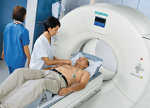
Siemens recently introduced its SOMATOM Definition Flash, which reportedly performs a cardiac CT scan in 1 mSv.
Fewer topics in computed tomography (CT) are hotter today than lowering radiation dose. From mainstream media articles on radiation exposure to conference sessions on dose-lowering techniques to new ultra-fast systems designed to reduce exposure, healthcare has put a premium on this issue. Accordingly, clinicians and manufacturers have approached the problem of radiation exposure in a variety of ways, while trying not to compromise image quality.
Cardiac CT
Perhaps no other CT scan has garnered more attention to dose reduction than cardiac CT. Last year, Toshiba America Medical Systems released its Aquilion One 320-detector row CT, designed to image the whole heart in a single heartbeat, thus exposing the patient to less radiation.
At RSNA 2008, Siemens Healthcare debuted its SOMATOM Definition Flash dual-source CT, a scanner touted as being able to scan the entire chest region in 0.6 seconds, while keeping the radiation dose lower than other scans. It is said that the Flash can perform a spiral heart scan at less than 1 millisevert (mSv), whereas the average effective dose required for this purpose usually ranges from 8 mSv to 40 mSv, according to the company. Siemens said this opens up the possibility for the Flash to do routine cardiac scans and help coronary CTA become the gold standard scan for patients with low-and-intermediate chest pain.
“That is the goal,” Bernd Montag, Ph.D., CEO of Siemens Healthcare Imaging & IT division, told Imaging Technology News at RSNA 2008. “To become the gold standard, it needs to work always. It also needs to be a safe exam.”
There are other methods for lowering cardiac CT dose. On GE Healthcare’s LightSpeed CT750, the scanner allows for modulating the kilovolts (kV) for 100 kV tube imaging. The scanner also uses the step-and-shoot mode, an imaging technique that limits exposure only to the portion of the cardiac cycle when data acquisition is relevant.
The step-and-shoot has significantly lowered the dose, according to James Min, M.D., associate professor of medicine, Weill Cornell Medical College at Cornell University, assistant attending physician, New York-Presbyterian Hospital in Ithaca, NY.
“We probably use it on about ninety percent of our patients and they are getting radiation doses of about 3 to 4 milliseverts routinely,” said Dr. Min. “It has probably reduced [radiation] by about 75 percent.”
The only patient population that clinicians can’t use the step-and-shoot method on is patients with high heart rates, according to Dr. Min. In those instances, the image quality is poor.
Sweden-based Sapheneia – an imaging hardware and software company – manufactures the Clarity CT Solution that aims to lower radiation dose by utilizing proprietary post-processing algorithms, enabling radiologists to use less radiation during the acquisition. The Cardiology Associates of Martin & SF in Larkspur, CA, recently began use the Clarity CT Solution during cardiac scans. According to James Adams, M.D., the radiation dose in coronary CTA scans and calcium scoring scans have decreased by over 50 percent.
Thoracic CT
At a refresher course at RSNA 2008, Narinder Paul, M.D., of the University of Toronto and Princess Margaret Hospital, Toronto, Ontario, Canada, demonstrated several cardiothoracic CT images (in non-pediatric cases) taken at different kilovolt peaks (kVp) to illustrate that using a lower kVp in many cases won’t compromise the integrity of the diagnosis, yet will significantly lower the radiation.
“The images might not be pretty – there may be some noise – but you can make the diagnosis,” Dr. Paul told the audience. In order to lower the kVp for CT scans, “your radiologists must buy into it.”
Dr. Paul noted that in some cases where multiple CT scans are needed for the same body part, it becomes particularly important to lower the dose.
Massachusetts General Hospital in Boston presented research at RSNA 2008 on studies they conducted using SharpView CT software (made by Sweden-based SharpView AB), an adaptive filter which constitutes mathematical algorithms that resemble the human vision system. The filter aims to reduce noise and improve image quality. The software is integrated in the image flow and the filtrated images are delivered automatically into the ordinary picture archive of the clinic. One of the hospital’s studies found that using the image filter software helped reduce radiation dose by as much as 75 percent without compromising diagnostic acceptability or lesion detection for chest multidectector CT scans.
Pediatric CT
There are a variety of issues in imaging pediatric patients. Deciding whether a CT (or any scan) is an important first step, according to Donald Frush, M.D., professor of radiology at the Duke University School of Medicine and chief of pediatric radiology at Duke Children’s Hospital and Health Center, and a leading pediatric radiation dose expert. Clinicians may be more apt to order a scan for children because they (the children) have less clinical history, and because clinicians may feel more anxiety to not miss anything when trying to diagnose a child.
Radiation is a big concern in pediatric imaging for a variety of reasons. Children are two to three times more sensitive to radiation in terms of biological effect, with some experts estimating children are up to 10 times more sensitive, according to Dr. Frush.
“You have to realize with kids that they might be getting a CT scan or two when they are five years old for some medical condition. One or two CT exams isn’t really going to make significant difference in terms of developing cancer, but you don’t know what that child is going to need at 10 or 15 or 20 or 30,” Dr. Frush said. “They may develop kidney stones and get five or 10 CTs. They have this cumulative possibility that someone who is 75 or 80 doesn’t.”
Dr. Frush developed protocols for CT radiation dose based on the Broselow-Luten system that has been used in emergency departments. The Color Coding for Kids CT protocols, found on GE Healthcare’s CT scanners, aim to lower the radiation dose in children by adjusting the parameters according to a patient’s size and weight.
“That’s what technologists and radiologists want,” Dr. Frush said. “They want to have size-appropriate protocols. Many of the previous protocols were based on age. Age is only a rough indicator of size.”
Dr. Frush believes that more can be done to lower the dose, including having better dose estimates on CT scanners for children, which is currently being worked on by The Alliance for Radiation Safety in Pediatric Imaging and other organizations.
Dr. Frush would like to see all vendors adopt a universal language that represents dose on the exam, and that the dose is not “buried somewhere in DICOM information” but is conveyed in a way that is easily understood.
Having protocol warnings on the scanner would also be helpful, said Dr. Frush, so if a protocol were put into the scanner that falls outside of a guideline range, a manual override would need to be made before the scan started.
“Right now you can do whatever you want and get whatever exam you want,” Dr. Frush said.
Trauma CT
Dose considerations for trauma victims are important since many victims are relatively young, according to Monique Brink, M.D., and Frank de Lange, Ph.D., of the department of diagnostic imaging at Radbound University Nijmegen Medical Centre in the Netherlands. They recently completed a clinical trial measuring how arm position affects the image quality and effective dose for CT scans of trauma patients using a 16-slice CT.
The study showed that positioning the patients’ arms above the shoulder, when possible, increased image quality and kept the median effective dose to 18.6 mSv. Patients positioned with one arm below the shoulder saw an 18 percent increase in radiation dose, while patients with both arms below the shoulder saw a 45 percent increase in dose. The substantial increase in mSv surprised Dr. Brink and de Lange.
According to the clinicians, AEC (automated exposure controlled) techniques automatically adapt the tube current to the patient’s attenuation and shape. In a scan of a patient’s torso, the arms contribute to a higher patient volume, thereby increasing radiation dose.
“We always have been using a protocol in which arm raising is preferred in trauma CT scanning,” Dr. Brink said. “However, after this study, not only radiologists and technicians, but also other trauma team members — surgeons, emergency physicians, anesthesiologists and nurses — are more aware to position the patients’ arms above the shoulder region.”
Maximize Awareness
Part of the challenge facing the medical community is training doctors how to apply the techniques to lower radiation exposure. The Society of Cardiovascular Computed Tomography (SCCT) serves as a tremendous educational resource for physicians and technologists, and is dedicated to ensuring expert and appropriate application of SCCT through training and accreditation for healthcare professionals. The society’s president-elect Jack Ziffer, Ph.D., M.D., who is chief of radiology at Baptist Hospital of Miami and the director of cardiac imaging at the Baptist Cardiac and Vascular Institute in Miami, points out that “dramatic dose reductions can be achieved when physicians and technologists are aware of what the doses actually are.”
He continued, “An additional approach is encouraging participation in laboratory accreditation through ICACTL (as an example), and physician certification through CBCCT, as mechanisms for referring physicians and patients to recognize laboratories and physicians that have documented expertise in CCTA and knowledge of dose reduction strategies.”
As the society’s new president, Dr. Ziffer says, “the SCCT will pursue efforts to ensure that awareness is maximized.”

 June 18, 2026


