June 26, 2018 — People who smoke or have diabetes may be at increased risk of calcifications in a region of the brain crucial to memory, according to a new study published online in the journal Radiology.
Researchers have hypothesized that abnormal buildups of calcium, or calcifications, in the hippocampus may be related to vascular problems that could contribute to hippocampal atrophy and subsequent cognitive deterioration. However, published research on the association between hippocampal calcification and cognitive impairment is limited.
“We know that calcifications in the hippocampus are common, especially with increasing age,” said the study’s lead author, Esther J.M. de Brouwer, M.D., a geriatrician at the University Medical Center in Utrecht, the Netherlands. “However, we did not know if calcifications in the hippocampus related to cognitive function.”
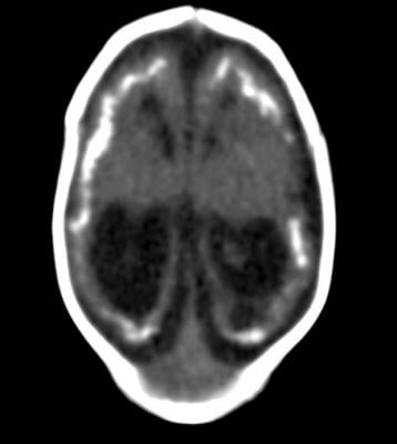
Advances in neuroimaging have provided opportunities to explore the role of hippocampal calcifications in dementia. The development of multiplanar brain computed tomography (CT) scans has enabled better distinction between hippocampal calcifications and calcifications in nearby brain structures like the choroid plexus.
“A multiplanar CT scan makes it possible to see the hippocampus in different anatomical planes; for example, from top to bottom, right to left, and front to back,” de Brouwer said. “Before multiplanar CT scans, hippocampal calcifications were often mistaken for choroid plexus calcifications. So with multiplanar CT scans, hippocampal calcifications are better distinguished from calcifications in other areas.”
de Brouwer and colleagues studied the association between vascular risk factors like high blood pressure, diabetes and smoking, and hippocampal calcifications. They also assessed the effects of calcifications on cognitive function.
The study group included 1,991 patients, average age 78 years, who had visited a memory clinic at a Dutch hospital between 2009 and 2015. The patients had a standard diagnostic workup including cognitive tests and brain CT scans. The researchers analyzed the CT scans for the presence and severity of hippocampal calcifications.
Of the 1,991 patients, 380 (19.1 percent) had hippocampal calcifications. Older age, diabetes and smoking were associated with an increased risk of hippocampal calcifications on CT scans.
While the study was not designed to conclusively determine if smoking and diabetes increase the risk of hippocampal calcifications, the results strongly suggest a link.
“We do think that smoking and diabetes are risk factors,” de Brouwer said. “In a recent histopathology study, hippocampal calcifications were found to be a manifestation of vascular disease. It is well known that smoking and diabetes are risk factors for cardiovascular disease. It is, therefore, likely that smoking and diabetes are risk factors for hippocampal calcifications.”
There was no link between the presence and severity of hippocampal calcifications and cognitive function — a surprising finding, according to de Brouwer, with several possible explanations.
“The hippocampus is made up of different layers, and it is possible that the calcifications did not damage the hippocampal structure that is important for memory storage,” she said. “Another explanation could be the selection of our study participants, who all came from a memory clinic.”
The researchers plan to carry out additional studies in different groups of people to better understand possible links between these calcifications and cognitive problems.
For more information: www.pubs.rsna.org/journal/radiology
Reference
de Brouwer E.J.M., Kockelkoren R., Claus J.J., et al. "Hippocampal Calcifications: Risk Factors and Association with Cognitive Function.” Radiology, June 12, 2018. https://doi.org/10.1148/radiol.2018172588

 June 18, 2026


