November 2, 2017 — Young hockey players who have suffered concussions may still show changes in the white matter of the brain months after being cleared to return to play, researchers at Western University have found. These findings were achieved through use of sophisticated magnetic resonance imaging (MRI) techniques.
The study, published in the Oct. 25, 2017, online issue of Neurology, the medical journal of the American Academy of Neurology, looked at MRI brain scans from 17 Bantam-level hockey players between the ages of 11 and 14, who suffered a concussion during the regular season and who were compared to an age-matched control of non-concussed players.
The athletes underwent brain MRI testing within 24-72 hours of the initial concussion, and again three months post-concussion, at which time all players reported no symptoms on clinical evaluations and were cleared to return to play following the standard concussion consensus Return to Play protocol. Most of the concussions were a result of falls that resulted in a hit to the back of the head.
"What the MRI shows is that there are still changes occurring in the brain even after the clinical tests have returned to normal," said Ravi Menon, Ph.D., professor at Western's Schulich School of Medicine & Dentistry and a scientist at Robarts Research Institute. "This is potentially of some concern and we'd like to understand this further to determine if these are normal healthy changes or if they are indicative of something that might be going wrong."
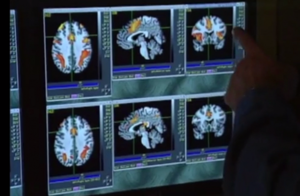
The advanced MRI data was analyzed by Ph.D. candidate Kathryn Manning at Western's Centre for Functional and Metabolic Mapping at Robarts Research Institute. The team looked at diffusion, functional and spectroscopy MRI data. On both the acute and the three-month scans, researchers observed that the very long fiber tracks in the brains of the concussed players were damaged, and also saw 'hyper-connectivity' in some areas of the brain, suggesting the brain may still have been trying to compensate for the injury.
"We saw that there were prolonged abnormalities in terms of the white matter in the brain," said Manning, noting that these changes are only visible using high field-strength MRI and these sophisticated analytical methods. "On a normal clinical MRI scan, you typically see the structural images of the brain, and for a mild brain injury like a concussion, we aren't able to see the underlying changes we were able to see using these advanced methods."
Lisa Fischer, M.D., assistant professor at Schulich Medicine & Dentistry, treats concussions at The Fowler Kennedy Sport Medicine Clinic, supported by Western University and London Health Sciences Centre. For her, this news is promising for concussion diagnosis. "If we can come up with a clinically-relevant, objective measure for concussion diagnosis and recovery, we can make safer decisions about return to play," she said. "This study has the potential to help develop that."
For more information: www.neurology.org

 July 02, 2026


