September 12, 2017 — Hoag Memorial Hospital Presbyterian recently became the first hospital in Orange County, Calif., to install the Siemens Somatom Force computed tomography (CT) scanner. The Somatom Force allows radiologists to obtain higher-quality images faster, more efficiently and with less radiation than older-generation CT scanners.
“This technology definitely raises the bar when it comes to performing detailed, high-resolution non-invasive cardiac imaging,” said radiologist Humberto Wong, M.D. “Among other things, the Force significantly improves the process for rapidly evaluating our patients with acute chest pain, improving accuracy, and, most importantly minimizing their radiation exposure.”
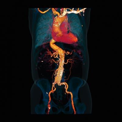
The scanner is capable of performing a coronary artery study, as well as an entire computed tomography angiogram (CTA) of the chest with a single injection of intravenous contrast, reducing the amount of radiation and volume of contrast needed, as well as the testing time, for patients who require both studies. The combination study allows for assessment of potentially blocked arteries to the heart, as well as pulmonary embolism or dissection of the aorta.
To gain experience with the new scanner, several Hoag physicians personally underwent the procedure; doctors on both sides of the scanner – testers and subjects. They say the scanner is truly an advanced imaging solution ideal for seniors and other patients for whom time and invasiveness are paramount issues.
Subbarao Myla, M.D., FACC, medical director of the Cardiac and Endovascular Labs and Cardiovascular Research at the Jeffrey M. Carlton Heart and Vascular Institute at Hoag, summarized the benefits best, saying, “The acquisition of this scanner is proof of Hoag’s commitment to high quality, cutting-edge technology and our patients. The speed of imaging will benefit our patients, while the quality and clarity of coronary images will reassure the physicians caring for chest pain patients. This is a major step forward in the low risk chest pain pathway.”
Perhaps most notably, Hoag clinicians have already learned it will reduce unnecessary hospitalizations and patient medical costs. Three days after Hoag installed the technology, a patient came to the emergency department with acute chest pain. Within one hour, images from the scanner excluded a heart attack and the patient was discharged that same evening instead of requiring a hospital admission.
“With the aid of this new technology, we are able to help our emergency department and cardiology colleagues avoid needless patient hospitalizations,” Wong said. “Instead of admitting the patient overnight for observation and additional tests, the ER physician had results within one hour after ordering the study and was able to safely discharge the patient with confidence that there was no acute coronary abnormality.”
An additional clinical benefit of the scanner’s speed for heart studies is that it negates the need for patients with rapid heart rates to take drugs, which slow the heartbeat to decrease motion blurring. With standard CT scanners, a heart rate of more than 70 beats per minute (bpm) makes it difficult to create sharp motion-free images, so doctors will often prescribe or administer medications (oral beta-blockers) to reduce the heart rate. Such drugs require at least one hour to take effect, introducing considerable delay, and add cost in the process.
“The Force can handle higher heart rates with ease,” Wong explained. “During our initial two weeks of testing, we performed more than 30 coronary CTAs, with most patients having heart rates over 70 beats per minute and some as high as 105 beats per minute and in all cases, the images were diagnostic.”
Besides its uses in cardiac imaging, the scanner also has improved speed and visualization of brain structures, including the arteries in the brain, which aids in rapid diagnosis, management and rescue of acute stroke patients. Studies of other organs and their vascular supply likewise benefit from the scanner’s capabilities.
For more information: www.usa.healthcare.siemens.com

 October 07, 2026