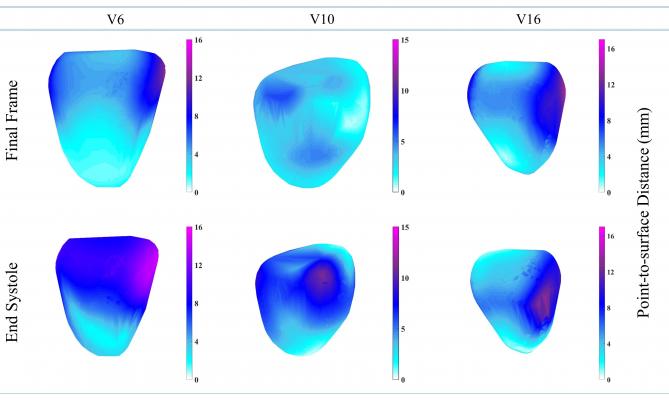
Displacement comparison at the end-systolic frame and final frame. The three patients (V6, V10, V16) with different left-ventricle walls are shown. Point-to-surface distance is a measure to estimate the distance of a point from the reference surface. Image courtesy of WMG, University of Warwick
August 28, 2019 — A new 3-D magnetic resonance imaging (MRI) computing technique developed by scientists in WMG at the University of Warwick focuses on hierarchical template matching (HTM) to diagnose cardiac disease without the use of gadolinium contrast. The technique is explored in an article in the journal Scientific Reports.1
MRI has long been used to diagnose cardiomyopathy, heart attacks, irregular heartbeats and other heart disease. Traditionally when a patient goes for an MRI scan they are given a dose of gadolinium, which reacts with the magnetic field of the scanner to produce an image of the protons in the metal realigning with the field. The faster the protons realign, the brighter the image features to show where the dead muscles are in the heart and what the diagnosis is.
The dose of gadolinium, however, can have detrimental effects to other parts of the body, particularly the risk of kidney failure.
The hierarchical template matching technique involves:
-
A numerically stable technique of left ventricular (LV) myocardial tracking;
-
A 3-D extension of local weighted mean function to transform MRI pixels; and
-
A 3-D extension of the HTM model for myocardial tracking problems.
Use of this technique eliminates the need for gadolinium, reducing the risk of damage to other organs.
Prof. Mark Williams from WMG at the University of Warwick said, “Using 3D MRI computing technique we can see in more depth what is happening to the heart, more precisely to each heart muscle, and diagnose any issues such as remodeling of the heart that causes heart failure. The new method avoids the risk of damaging the kidney opposite to what traditional methods do by using gadolinium.”
Jayendra Bhalodiya, who conducted the research from WMG, University of Warwick added, “This new MRI technique also takes away stress from the patient, as during an MRI the patient must be very still in a very enclosed environment, meaning some people suffer from claustrophobia and have to stop the scan. Often when they do this they have to administer another dose of the damaging gadolinium and start again. This technique doesn’t require a dosage of anything, as it tracks the heart naturally.”
For more information: www.nature.com
Related Content
FDA Approves Bayer's Gadavist Contrast for Cardiac MRI in Adult Coronary Artery Disease Patients
Using Artificial Intelligence to Reduce Gadolinium Contrast
The Debate Over Gadolinium MRI Contrast Toxicity
Reference
1. Bhalodiya J.M., Palit A., Ferrante E., et al. Hierarchical Template Matching for 3D Myocardial Tracking and Cardiac Strain Estimation. Nature Scientific Reports, published online Aug. 28, 2019. https://doi.org/10.1038/s41598-019-48927-2

 July 10, 2026