August 11, 2015 — Brainreader, a Danish medical device company, announced the launch of its new software product that has the potential to speed the detection of changes in a patient's brain volume. These are a key indicator in the diagnosis of degenerative brain diseases such as Alzheimer's disease, epilepsy and depression.
The software, Neuroreader, also can help identify changes in brain volume associated with concussions and other traumatic brain injuries.
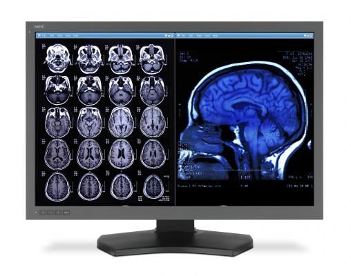
In use by a growing number of radiologists and neuroradiologists in the United States and Europe, Neuroreader analyzes magnetic resonance imaging (MRI) scans of patients' brains and benchmarks them against a U.S. Food & Drug Administration (FDA)-cleared database of healthy brain tissues in a matter of minutes. The software measures 45 specific structures within the brain.
"Brain scans are typically analyzed visually by radiologists in what can be a time-consuming process, especially when changes in brain volume are slight," said Jamila Ahdidan, Ph.D., founder and chief technology officer of Brainreader. "When looking for changes as small as hundredths of a millimeter, a software-driven analysis allows for these tiny changes to be detected in minutes."
For more than 30 years, since the development of the MRI scanner, physicians have ordered MRI scans of the brain to identify traumatized, diseased or atrophied tissue.
"Radiologists rely on our human pattern recognition to identify tiny abnormalities in patients' brains," said Barton Branstetter, M.D., neuroradiologist at University of Pittsburgh Medical Center, Presbyterian. "But even the best radiologist cannot precisely determine the volume of every structure in the brain without hours of intensive manual calculations. Neuroreader gives us easy, reliable, reproducible volume measurements and takes the guesswork out of analyzing structures in our patients' brains."
When treating a patient with signs of neurodegenerative diseases, the physician simply uploads the MR image to the Brainreader server. Neuroreader then analyzes the MRI scan and delivers a report in less than five minutes, indicating which brain structures' volume are abnormal and to what extent. The software, which has an intuitive interface, produces an easily interpreted automated report.
One of the brain structures analyzed is the hippocampus, the measurement of which is vital in determining a person's ability to maintain strong cognitive function, according to neurologist and Alzheimer's disease expert Majid Fotuhi, M.D., Ph.D., chairman, NeuroGrow Brain Fitness Center, McLean, Virginia, and Johns Hopkins Medicine, Baltimore.
"Measuring the size of the hippocampus is the single most important factor in determining a person's prognosis for having strong memory performance in the future," said Fotuhi. "That's where the Neuroreader comes in. It's a major breakthrough in establishing an accurate measurement of this part of the brain."
According to Fotuhi, the Neuroreader will help him and his colleagues diagnose patients early for degenerative brain diseases and to create treatment plans that can not only slow the loss of brain tissue, but even to build it back up. "In the future, I believe it will become routine for patients - and their doctors - to know the volume of their hippocampus and to have it monitored regularly," he said.
According to Dale E. Bredesen, M.D., Augustus Rose Professor of Neurology at UCLA, and founding president and CEO of the Buck Institute for Research on Aging: "Recent publications have offered some of the first evidence for functional, sustained improvement in neurodegenerative conditions, and the ability to quantitate multiple brain region volumes reproducibly and longitudinally will be critical as therapeutic programs are being refined. Neuroreader is perfectly suited for this."
Neuroreader, which is sold as software-as-a-service, is available to hospitals radiology and neuroradiology departments on a pay-per-use or subscription model tailored to the healthcare organization's wishes.
For more information: www.brainreader.net

 July 10, 2026


