News | Magnetic Resonance Imaging (MRI) | June 07, 2019

Competition had leading academics evaluate MRI, PET, CT and combination images from MR Solutions users around the world

A high-fidelity 3-D tractography of the left ventricle heart muscle fibers of a mouse

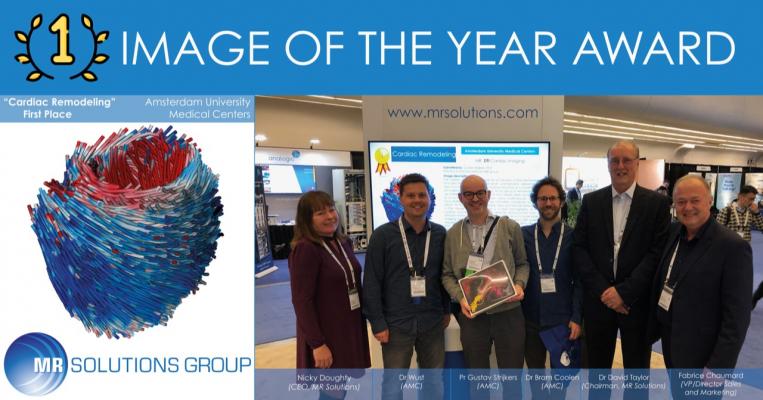
Figure 1. A high-fidelity 3-D tractography of the left ventricle heart muscle fibers of a mouse from Amsterdam Ph.D. researcher Gustav Strijkers.


June 7, 2019 — The Amsterdam University Medical Center has won MR Solutions’ Image of the Year 2019 award for the best molecular research image. Preclinical image submissions came in from most of MR Solutions’ users across the world. The winners, which were presented at the International Society for Magnetic Resonance in Medicine (ISMRM) conference, May 11-16 in Montreal, QC, Canada, were chosen by a panel of leading academics who examined images from magnetic resonance imaging (MRI), positron emission tomography (PET) or computed tomography (CT) – or a combination of imaging modalities.

The winning submission was carried out by Amsterdam Ph.D. researcher Gustav Strijkers. This technique was used in cardiac research to produce a high-fidelity 3-D tractography of the left ventricle heart muscle fibers of a mouse (figure 1). The exceptional quality and accuracy of the cardiomyopathy research significantly advances the preclinical imaging sector in the battle to better understand heart muscle disease and treatment.

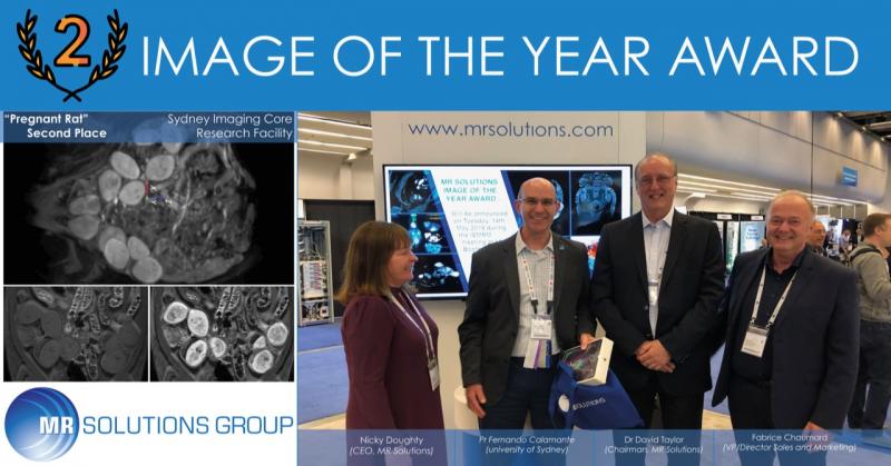
The submission from Sydney Imaging Core Research Facility came a close second place for the high-quality MR abdominal image of a pregnant Sprague Dawley rat (figure 2). The resulting image showed multiple embryonic implants and was ranked for image clarity, research interpretation and understanding.

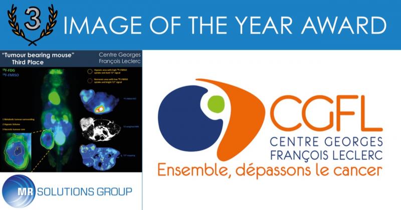
The third place submission by the Centre Georges François Leclerc centre in France was a PET-MR whole body image of an eight-week-old BALB/c mouse showing a CT26 xenograft tumor (figure 3). This displayed a clear breakdown of the metabolic, hypoxic and necrotic regions of the tumor. Additional axial images showed further localized information on the high and low uptake areas of 18F-FMISO using a 2T signal.

For more information: www.mrsolutions.com


Related Content

News | Contrast Agents

April 23, 2026 — On April 23, GE HealthCare announced the first patient has been dosed in the international, multi ...

Time April 23, 2026
arrow
News | Radiology Imaging

April 20, 2026 — Bracco Imaging has announced a strategic alliance with NYU Langone Health to advance innovation in ...

Time April 23, 2026
arrow
News | Radiology Imaging

April 7, 2026 — Onvida Health and Siemens Healthineers have entered a 10-year Value Partnership¹ designed to bring the ...

Time April 09, 2026
arrow
News | SNMMI

The Society of Nuclear Medicine and Molecular Imaging's (SNMMI) 2026 Annual Meeting will take place May 30–June 2 in Los ...

Time April 07, 2026
arrow
News | PET Imaging

March 25, 2026 — Catalyst MedTech, a provider of nuclear medicine and molecular imaging solutions, has announced it is ...

Time April 06, 2026
arrow
News | Radiology Business

March 31, 2026 — Radon Medical Imaging, a medical imaging equipment maintenance and repair services company, has has ...

Time March 31, 2026
arrow
News | Radiology Imaging

March 26, 2026 — GE HealthCare has announced a renewed research collaboration with Stanford Medicine Department of ...

Time March 30, 2026
arrow
News | Magnetic Resonance Imaging (MRI)

March 25, 2026 A Penn Medicine–led team has developed a first‑of‑its‑kind artificial intelligence system that interprets ...

Time March 26, 2026
arrow
News | Radiology Education

March 17, 2026 – The Center for Radiology Education (CRE) has announced a nationwide initiative to provide scholarships* ...

Time March 17, 2026
arrow
News | Radiology Business

March 12, 2026 — DelveInsight's has released its latest Diagnostic Imaging Equipment Market Insights report. The in ...

Time March 13, 2026
arrow
Subscribe Now